Project Week 25/Slicer and 3D Printing

From NAMIC Wiki
Jump to: navigation, search
Home < Project Week 25 < Slicer and 3D Printing


Back to Projects List

Key Investigators

Project Description

From DICOM data to a 3D print of anatomical models for training: anatomy classes and/or surgical planning

Objective Approach and Plan Progress and Next Steps
  • Develop and document a workflow to process the acquired DICOM images
  • Improve the segmentation mechanics in order to extract the data to print
  • Prepare the STL files for printing: checking the mesh and creating the support structures
  • Before Project Week:
    • Know the "state of the art" of 3d printer options and materials
    • Diving into the DICOM and NRRD file structure
  • During Project Week:
    • Establish the guidelines for the segmentation procedures
    • Identify the STL export issues
  • Progress
    • We've clarified the segmentation procedure using the new segmentation editor on the last Slicer version
    • Also we've gotten some fresh information about prototypes materials used by other groups in order to create their lifelike models and phantoms (gellatins, silicons, plastics and such)
  • Next step
    • Start printing our first phantom prototype parts and building the rest of the needed parts with the other complementary materials
    • Stablish a workflow for segmentation, as generalistic as possible in order to make it applicable for as many different segmentation needs as possible (focusing on how you use the segmentation editor, not on the actual anatomical knowledge or the different DICOM sources availables)

Illustrations

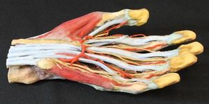
3D printed model (hand colored)
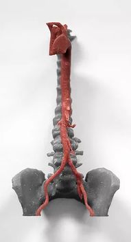
3D print of a human aorta

Background and References

Possible application areas / IDEAS

  • Life size models of different anatomical structures: normal and pathological
  • Create life like models, that provide a sensorial feeling as close as the "organic" have