Projects:InteractiveSegmentation
Back to Boston University Algorithms
Interactive Image Segmentation With Active Contours
A driving clinical study for the present work is a population study of skeletal development in youth. Bone grows from the physis (growth plate), located in the middle of a long bone between the epiphysis and metaphysis. Full adult growth is reached when the physis disappears completely. Precise understanding of how the growth plates in femur and tibia change from childhood to adulthood enables improved surgical planning(e.g. determining tunnel placement for anterior cruciate ligament (ACL) reconstruction) to avoid stunting the patient's growth or compromising the stability of the knee. Currently, growth potential is measured by a physician using an x-ray scan of multiple bones; patients receive repeated doses of radiation exposure.
Description
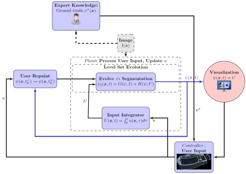
In this work, interactive segmentation is integrated with an active contour model and segmentation is posed as a human-supervisory control problem. User input is tightly coupled with an automatic segmentation algorithm leveraging the user's high-level anatomical knowledge and the automated method's speed. Real-time visualization enables the user to quickly identify and correct the result in a sub-domain where the variational model's statistical assumptions do not agree with his expert knowledge. Methods developed in this work are applied to magnetic resonance imaging (MRI) volumes as part of a population study of human skeletal development. Segmentation time is reduced by approximately five� times over similarly accurate manual segmentation of large bone structures.
Flowchart for the interactive segmentation approach. Notice the user's pivotal role in the process.
Time-line of user input into the system. Note that user input is sparse, has local effect only, and decreases in frequency and magnitude over time.
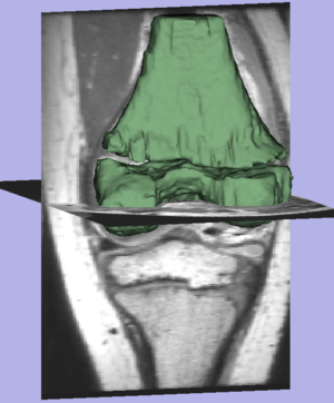
Result of the segmentation.
Current State of Work
We have gathered a sample data set; currently, we are registering the scan to create a united patient model. This model will localize structures and constrain shape. Additionally, we are investigation interaction approaches for registration and segmentation that will "put the user in the loop" but greatly reduce the work compared to manual approaches.
Key Investigators
- Georgia Tech: Ivan Kolesov, Peter Karasev, Karol Chudy, and Allen Tannenbaum
- Emory University: Grant Muller and John Xerogeanes
Publications
In Press
I. Kolesov, P.Karasev, G.Muller, K.Chudy, J.Xerogeanes, and A. Tannenbaum. Human Supervisory Control Framework for Interactive Medical Image Segmentation. MICCAI Workshop on Computational Biomechanics for Medicine 2011.
P.Karasev, I.Kolesov, K.Chudy, G.Muller, J.Xerogeanes, and A. Tannenbaum. Interactive MRI Segmentation with Controlled Active Vision. IEEE CDC-ECC 2011.


