Slicer:Feedback
3D Slicer is a free open source software package distributed under a BSD style license. The majority of funding for the development of 3D slicer comes from a number of grants and contracts from the National Institutes of Health (see slicer acknowledgements for more information).
We invite you to provide information on how you are using 3D Slicer to produce peer-reviewed research. Information about the scientific impact of this tool is helpful in raising funding for the continued support of this tool.
Please email us or add your paper or grant information to the bottom of this page. (Note: adding information here is easy - just create an account using the link at the top of this page, and then send your wiki username to namic-wiki@na-mic.org asking for permission to add links to the wiki. See the Editing Guide for more information.)
3D Slicer Enabled Research
Contents
- 1 3D Slicer Enabled Research
- 1.1 Using 3D Slicer in Astronomy
- 1.2 Registered, Sensor-Integrated Virtual Reality for Surgical Applications
- 1.3 A 3D Model Simulating Sediment Transport, Erosion and Deposition within a Network of Channel Belts and an Associated Floodplain
- 1.4 Appearance of the Levator Ani Muscle Subdivisions in Magnetic Resonance Images
- 1.5 2D Rigid Registration of MR Scans using the 1D Binary Projections
- 1.6 Habitual Use of the Primate Forelimb is Reflected in the Material Properties of Subchondral Bone in the Distal Radius
- 1.7 Molecular Diffusion in MRI: Technical Application of Fiber Tracking
- 1.8 Range of Curvilinear Distraction Devices Required for Treatment of Mandibular Deformities
- 1.9 Developmental Response to Cold Stress in Cranial Morphology of Rattus: Implications for the Interpretation of Climatic Adaptation in Fossil Hominins
- 1.10 Abnormal Association between Reduced Magnetic Mismatch Field to Speech Sounds and Smaller Left Planum Temporale Volume in Schizophrenia
- 1.11 Quantification of Levator Ani Cross-sectional Area Differences between Women with and those without Prolapse
- 1.12 Vaginal Thickness, Cross-Sectional Area, and Perimeter in Women with and Those without Prolapse
- 1.13 Magnetic Resonance Imaging and 3-Dimensional Analysis of External Anal Sphincter Anatomy
- 1.14 Measurement of the Pubic Portion of the Levator ani Muscle in Women with Unilateral Defects in 3D Models from MR Images
- 1.15 A Comparison of Upper Airway Structures in Male and Female Obstructive Sleep Apnoea (OSA) Patients
- 1.16 Atlas Guided Identification of Brain Structures by Combining 3D Segmentation and SVM Classification
- 1.17 Development of a CAD (Computer Assisted Detection) System to Detect Lung Nodules in CT Scans
- 1.18 Dynamic Simulation of Joints Using Multi-Scale Modeling
- 1.19 Quasi-isometric Flattening of Curved Surfaces for Medical Imaging
- 1.20 3D Multiscale Agent-Based Cancer Model Using MR-Imaging Data
- 1.21 A Translation Station for Imaging
- 1.22 Preliminary Study on Digitized Nasal and Temporal Bone Anatomy
- 1.23 Group-Slicer: a collaborative extension of 3D-Slicer
- 1.24 Open-configuration MR-guided Microwave Thermocoagulation Therapy for Metastatic Liver Tumors from Breast Cancer
- 1.25 Virtual Cystoscopy - A Surgical Planning and Guidance Tool
- 1.26 Morphology, constraints, and scaling of frontal sinuses in the hartebeest, Alcelaphus buselaphus (Mammalia: Artiodactlya, Bovidae)
- 1.27 A ceratopsid dinosaur parietal from New Mexico and its implications for ceratopsid biogeography and systematics
Using 3D Slicer in Astronomy
Institution: The Initiative in Innovative Computing (IIC), Harvard University, Cambridge, Massachusetts
Project: IIC AstroMed Project
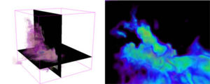
Background/Purpose: While astronomy and medical imaging seem very different, both fields search large amounts of image data looking for meaningful patterns. For example, a physician may inspect a patient's MRI scans looking for signs of disease, while an astronomer will analyze radio telescope image data to find evidence of a new star being born. The two sciences have separately developed many techniques to analyze, visualize, and catalog complex multi-dimensional imaging data, but seldom have experts from the two areas worked together.
The 3D Slicer software package is AstroMed's core tool for astronomical 3D data visualization. Originally designed for medical image analysis, 3D Slicer has many capabilities that are uncommon in astronomy software.
The AstroMed project brings together researchers from the Harvard Medical School and the Harvard-Smithsonian Center for Astrophysics, along with both national and international collaborators, to combine their knowledge and advance the state-of-the-art in both medical imaging and astronomy.
Registered, Sensor-Integrated Virtual Reality for Surgical Applications
Publication: IEEE Virtual Reality Conference 2007; PDF
Authors: Brady W. King, Luke A. Reisner, Michael D. Klein, Gregory W. Auner, Abhilash K. Pandya
Institution: Wayne State University, Detroit, Michigan, USA
Background/Purpose: The visualization for our image-guided surgery system is implemented using 3D Slicer, an open-source application for displaying medical data. 3D Slicer provides a virtual reality environment in which various imaging modalities (e.g. CT or MRI data) can be presented. The software includes the ability to display the locations of objects with respect to 3D models that are derived from segmentation of the medical imaging.
We modified 3D Slicer in several ways to adapt it to our application. First, we developed a TCP/IP interface that receives the tracking data for the MicroScribe and displays its position in the VR environment relative to the medical imaging data. This allows us to track the Raman probe in real-time. Second, we developed a way to place colored markers that indicate tissue/material classification on the medical imaging data. The combination of these modifications enables us to denote the location and classification of tissue/material scanned with the probe in near-real-time.
A 3D Model Simulating Sediment Transport, Erosion and Deposition within a Network of Channel Belts and an Associated Floodplain
Publication: PowerPoint Presentation PDF
Authors: Derek Karssenberg and John Bridge
Institution: Utrecht University, the Netherlands and Binghamton University, NY, USA.
Appearance of the Levator Ani Muscle Subdivisions in Magnetic Resonance Images
Publication: Obstet Gynecol. 2006 May ; 107(5): 1064–1069. PDF
Authors: Rebecca U. Margulies, Md, Yvonne Hsu, Md, Rohna Kearney, Mrcog, Tamara Stein, Phd, Wolfgang H. Umek, Md, and John O. L. DeLancey, Md
Institution: University of Michigan, Ann Arbor, Michigan; University College Hospital, London, United Kingdom; and Medical University Vienna, Vienna, Austria.
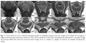
Background/Purpose: Identify and describe the separate appearance of 5 levator ani muscle subdivisions seen in axial, coronal, and sagittal magnetic resonance imaging (MRI) scan planes. METHODS: Magnetic resonance scans of 80 nulliparous women with normal pelvic support were evaluated. Characteristic features of each Terminologia Anatomica–listed levator ani component were determined for each scan plane. Muscle component visibility was based on pre- established criteria in axial, coronal, and sagittal scan planes: 1) clear and consistently visible separation or 2) different origin or insertion. Visibility of each of the levator ani subdivisions in each scan plane was assessed in 25 nulliparous women.RESULTS: In the axial plane, the puborectal muscle can be seen lateral to the pubovisceral muscle and decussating dorsal to the rectum. The course of the puboperineal muscle near the perineal body is visualized in the axial plane. The coronal view is perpendicular to the fiber direction of the puborectal and pubovisceral muscles and shows them as “clusters” of muscle on either side of the vagina. The sagittal plane consistently demonstrates the puborectal muscle passing dorsal to the rectum to form a sling that can consistently be seen as a “bump.” This plane is also parallel to the pubovisceral muscle fiber direction and shows the puboperineal muscle. CONCLUSION: The subdivisions of the levator ani muscle are visible in MRI scans, each with distinct morphology and characteristic features.
Grant Support:
- NICHD P50 HD 44406
- R01 HD 38665
2D Rigid Registration of MR Scans using the 1D Binary Projections
Publication: Enformatika Transactions on Engineering, Computing and Technology, November 2005; 9:157-161. PDF
Author: Panos D. Kotsas
Institution: Department of Automated Control and System Engineering, University of Sheffield, UK.
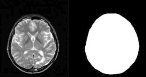
Background/Purpose: This research deals with the application of a signal intensity independent registration criterion for 2D rigid body registration of medical images using 1D binary projections. The criterion is defined as the weighted ratio of two projections. The ratio is computed on a pixel per pixel basis and weighting is performed by setting the ratios between one and zero pixels to a standard high value. The mean squared value of the weighted ratio is computed over the union of the one areas of the two projections and it is minimized using the Chebyshev polynomial approximation using n=5 points. The sum of x and y parallel projections is used for translational adjustment and a range of parallel projections between 40-50 deg (depending on the orientation of the image) for rotational adjustment. MR-MR registration experiments were performed and gave mean errors well below 1deg and 1 pixel (0.12 deg and 0.47 pixels for the example given in Enformatika Publication). The method is to be extended for surface matching.
Habitual Use of the Primate Forelimb is Reflected in the Material Properties of Subchondral Bone in the Distal Radius
Publication: J Anat. 2006 Jun; 208(6):659-670. PDF
Authors: Carlson KJ, Patel BA.
Institution: Department of Anatomical Sciences, School of Medicine, Stony Brook University, USA.
Background/Purpose: Bone mineral density is directly proportional to compressive strength, which affords an opportunity to estimate in vivo joint load history from the subchondral cortical plate of articular surfaces in isolated skeletal elements. Subchondral bone experiencing greater compressive loads should be of relatively greater density than subchondral bone experiencing less compressive loading. Distribution of the densest areas, either concentrated or diffuse, also may be influenced by the extent of habitual compressive loading. We evaluated subchondral bone in the distal radius of several primates whose locomotion could be characterized in one of three general ways (quadrupedal, suspensory or bipedal), each exemplifying a different manner of habitual forelimb loading (i.e. compression, tension or non-weight-bearing, respectively). We employed computed tomography osteoabsorptiometry (CT-OAM) to acquire optical densities from whichfalse-colour maps were constructed. The false-colour maps were used to evaluate patterns in subchondral density (i.e. apparent density). Suspensory apes and bipedal humans had both smaller percentage areas and less well-defined concentrations of regions of high apparent density relative to quadrupedal primates. Quadrupedal primates exhibited a positive allometric effect of articular surface size on high-density area, whereas suspensory primates exhibited an isometric effect and bipedal humans exhibited no significant relationship between the two. A significant difference between groups characterized by predominantly compressive forelimb loading regimes vs. tensile or non-weight-bearing regimes indicates that subchondral apparent density in the distal radial articular surface distinguishes modes of habitually supporting of body mass.
Grant Support: Funding for this research was provided by Sigma Xi Grants-In-Aid of Research to B.A.P.
Molecular Diffusion in MRI: Technical Application of Fiber Tracking
Publication: PowerPoint Presentation PPT
Author: Martha Liliana Mora V.
Institution: Rey Juan Carlos University, Madrid, Spain.
Range of Curvilinear Distraction Devices Required for Treatment of Mandibular Deformities
Publication: J Oral Maxillofac Surg. 2006 Feb;64(2):259-64.
Authors: Ritter L, Yeshwant K, Seldin EB, Kaban LB, Gateno J, Keeve E, Kikinis R, Troulis MJ
Institution: Department of Oral and Maxillofacial Surgery, Massachusetts General Hospital, Boston, MA, USA.
Background/Purpose: The purpose of this study was to determine the range of fixed trajectory curvilinear distraction devices required to correct a variety of severe mandibular deformities. MATERIALS AND METHODS: Preoperative computed tomography (CT) scans from 18 patients with mandibular deformities were imported into a CT-based software program (Osteoplan). Three-dimensional virtual models of the individual skulls were made with landmarks to track movements. An ideal treatment plan was created for each patient. Upper and lower boundaries for the dimensions of curvilinear distractors were established based on manufacturing and geometric constraints. Then, anatomically acceptable distractor attachment points were identified on the models using proximal and distal grids. Treatment plans were simulated for a series of distractors with varying radii of curvature, elongations (arc-length of device), and placements along the grids. The outcomes using these distractors were compared with the ideal treatment plans. Discrepancies were quantified in millimeters by comparing landmarks in the simulated versus ideal movements. RESULTS: Approximately 400,000 simulated 3-dimensional movements, based on the distractor parameters and variations in placement were computationally evaluated for the 18 cases. It was determined that, by varying distractor placement, a family of 5 distractors, with 3, 5, 7, and 10 cm radii of curvature and a straight-line device, could be used to treat all 18 cases to within 1.8 mm of error. CONCLUSIONS: The results of this study indicate that a family of 5 curvilinear distractors may suffice to treat a broad range of mandibular deformities.
Developmental Response to Cold Stress in Cranial Morphology of Rattus: Implications for the Interpretation of Climatic Adaptation in Fossil Hominins
Publication: Proc. R. Soc. B (2006) 273, 2605–2610. PDF
Authors: Todd C. Rae, Una Strand Viðarsdottir, Nathan Jeffery A. Theodore Steegmann Jr
Institution: Evolutionary Anthropology Research Group, Department of Anthropology, University of Durham, 43 Old Elvet, Durham DH1 3HN, UK
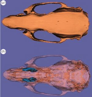
Background/Purpose: Adaptation to climate occupies a central position in biological anthropology. The demonstrable relationship between temperature and morphology in extant primates (including humans) forms the basis of the interpretation of the Pleistocene hominin Homo neanderthalensis as a cold-adapted species. There are contradictory signals, however, in the pattern of primate craniofacial changes associated with climatic conditions. To determine the direction and extent of craniofacial change associated with temperature, and to understand the proximate mechanisms underlying cold adaptations in vertebrates in general, dry crania rom previous experiments on cold- and warm-reared rats were investigated using computed tomography scanning and three-dimensional digitization of cranial landmarks. Aspects of internal and external cranial morphology were compared using standard statistical and geometric morphometric techniques. The results suggest that the developmental response to cold stress produces subtle but significant changes in facial shape, and a relative decrease in the volume of the maxillary sinuses (and nasal cavity), both of which are independent of the size of the skull or postcranium. These changes are consistent with comparative studies of temperate climate primates, but contradict previous interpretations of cranial morphology of Pleistocene Hominini.
Grant Support: None. Prof. Steegmann supplied the materials, and the time on the pQCT machine was graciously donated by Prof. Tim Skerry of the Royal Veterinary College, London.
Abnormal Association between Reduced Magnetic Mismatch Field to Speech Sounds and Smaller Left Planum Temporale Volume in Schizophrenia
Publication: NeuroImage (2004); 22:720–727. PDF
Authors: Hidenori Yamasue, Haruyasu Yamada, Masato Yumoto, Satoru Kamio, Noriko Kudo, Miki Uetsuki, Osamu Abe, Rin Fukuda, Shigeki Aoki, Kuni Ohtomo, Akira Iwanami Nobumasa Kato, Kiyoto Kasai
Institution: Department of Neuropsychiatry, Graduate School of Medicine, University of Tokyo, Bunkyo, Tokyo 113-8655, Japan
Background/Purpose: Schizophrenia is associated with language-related dysfunction. A previous study [Schizophr. Res. 59 (2003c) 159] has shown that this abnormality is present at the level of automatic discrimination of change in speech sounds, as revealed by magnetoencephalographic recording of auditory mismatch field in response to across-category change in vowels. Here, we investigated the neuroanatomical substrate for this physiological abnormality. Thirteen patients with schizophrenia and 19 matched control subjects were examined using magnetoencephalography (MEG) and high-resolution magnetic resonance imaging (MRI) to evaluate both mismatch field strengths in response to change between vowel /a/ and /o/, and gray matter volumes of Heschl's gyrus (HG) and planum temporale (PT). The magnetic global field power of mismatch response to change in phonemes showed a bilateral reduction in patients with schizophrenia. The gray matter volume of left planum temporale, but not right planum temporale or bilateral Heschl's gyrus, was significantly smaller in patients with schizophrenia compared with that in control subjects. Furthermore, the phonetic mismatch strength in the left hemisphere was significantly correlated with left planum temporale gray matter volume in patients with schizophrenia only. These results suggest that structural abnormalities of the planum temporale may underlie the functional abnormalities of fundamental language-related processing in schizophrenia.
Grant Support:
- C12670928 Ministry of Education, Culture, Sport, Science and Technology, Japan
- Welfide Medicinal Research Fundation, Japan
- Uehara Memorial Fundation, Japan
Quantification of Levator Ani Cross-sectional Area Differences between Women with and those without Prolapse
Publication: Obstet Gynecol. 2006 Oct;108(4):879-83. PDF
Authors: Hsu Y, Chen L, Huebner M, Ashton-Miller JA, DeLancey JO.
Institution: University of Michigan, Ann Arbor, Michigan, USA
Background/Purpose: Compare levator ani cross-sectional area as a function of prolapse and muscle defect status. METHODS: Thirty women with prolapse and 30 women with normal pelvic support were selected from an ongoing case-control study of prolapse. For each of the two groups, 10 women were selected from three categories of levator defect severity: none, minor, and major identified on supine magnetic resonance scans. Using those scans, three-dimensional (3D) models of the levator ani muscles were made using a modeling program (3D Slicer), and cross-sections of the pubic portion were calculated perpendicular to the muscle fiber direction using another program, I-DEAS. An analysis of variance was performed. RESULTS: The ventral component of the levator muscle of women with major defects had a 36% smaller cross-sectional area, and women with minor defects had a 29% smaller cross-sectional area compared with the women with no defects (P < .001). In the dorsal component, there were significant differences in cross-sectional area according to defect status (P = .03); women with major levator defects had the largest cross-sectional area compared with the other defect groups. For each defect severity category (none, minor, major), there were no significant differences in cross-sectional area between women with and those without prolapse. CONCLUSION: Women with visible levator ani defects on magnetic resonance imaging had significantly smaller cross-sectional areas in the ventral component of the pubic portion of the muscle compared with women with intact muscles. Women with major levator ani defects had larger cross-sectional areas in the dorsal component than women with minor or no defects.
Grant Support:
- NIH R01 HD-38665
- DGF HU1502/1-1
Vaginal Thickness, Cross-Sectional Area, and Perimeter in Women with and Those without Prolapse
Publication: Obstet Gynecol. 2005 May;105(5 Pt 1):1012-7. PDF
Authors: Hsu Y, Chen L, Delancey JO, Ashton-Miller JA
Institution: Division of Gynecology, Department of Obstetrics and Gynecology, University of Michigan Medical School, Ann Arbor, Michigan, USA.
Background/Purpose: Use axial magnetic resonance imaging to test the null hypothesis that no difference exists in apparent vaginal thickness between women with and those without prolapse. METHODS: Magnetic resonance imaging studies of 24 patients with prolapse at least 2 cm beyond the introitus were selected from an ongoing study comparing women with prolapse with normal control subjects. The magnetic resonance scans of 24 women with prolapse (cases) and 24 women without prolapse (controls) were selected from those of women of similar age, race, and parity. The magnetic resonance files were imported into an experimental modeling program, and 3-dimensional models of each vagina were created. The minimum transverse plane cross-sectional area, mid-sagittal plane diameter, and transverse plane perimeter of each vaginal model were calculated. RESULTS: Neither the mean age (cases 58.6 years +/- standard deviation [SD] 14.4 versus controls 59.4 years +/- SD 13.2) nor the mean body mass index (cases 24.1 kg/m(2)+/- SD 3.3, controls 25.7 kg/m(2)+/- SD 3.7) differed significantly between groups. Minimum mid-sagittal vaginal diameters did not differ between groups. Patients with prolapse had larger minimum vaginal cross-sectional areas than controls (5.71 cm(2)+/- standard error of the mean [SEM] 0.25 versus 4.76 cm(2)+/- SEM 0.20, respectively; P = .005). The perimeter of the vagina was also larger in the prolapse group (11.10 cm +/- SEM 0.24) compared with controls (9.96 cm +/- SEM 0.22) P = .001. Subgroup analysis of patients with endogenous or exogenous estrogen showed prolapse patients had larger vaginal cross-sectional area (P = .030); in patients without estrogen group differences were not significant (P = .099). CONCLUSION: Vaginal thickness is similar in women with and those without pelvic organ prolapse. The vaginal perimeter and cross-sectional areas are 11% and 20% larger in prolapse patients, respectively. Estrogen status did not affect differences found between groups.
Grant Support:
- NIH R01 HD038665
Magnetic Resonance Imaging and 3-Dimensional Analysis of External Anal Sphincter Anatomy
Publication: Obstet Gynecol. 2005 Dec;106(6):1259-65. PDF
Authors: Hsu Y, Fenner DE, Weadock WJ, DeLancey JO
Institution: Pelvic Floor Research Group, Division of Gynecology, Department of Obstetrics and Gynecology, University of Michigan, Ann Arbor, Michigan 48109-0276, USA.
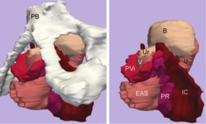
Background/Purpose: To use magnetic resonance images of living women and 3-dimensional modeling software to identify the component parts and characteristic features of the external anal sphincter (EAS) that have visible separation or varying origins and insertions. METHODS: Detailed structural analysis of anal sphincter anatomy was performed on 3 pelvic magnetic resonance imaging (MRI) data sets selected for image clarity from ongoing studies involving nulliparous women. The relationships of anal sphincter structures seen in axial, sagittal, and coronal planes were examined using the 3-D Slicer 2.1b1 software program. The following were requirements for sphincter elements to be considered separate: 1) a clear and consistently visible separation or 2) a different origin or insertion. The characteristic features identified in this way were then evaluated in images from an additional 50 nulliparas for the frequency of feature visibility. RESULTS: There were 3 components of the EAS that met criteria as being "separate" structures. The main body (EAS-M) is separated from the subcutaneous external anal sphincter (SQ-EAS) by a clear division that could be observed in all (100%) of the MRI scans reviewed. The wing-shaped end (EAS-W) has fibers that do not cross the midline ventrally, but have lateral origins near the ischiopubic ramus. This EAS-W component was visible in 76% of the nulliparas reviewed. CONCLUSION: Three distinct external anal sphincter components can be identified by MRI in the majority of nulliparous women.
Grant Support:
- P50 HD4406
- R01 HD38665
Measurement of the Pubic Portion of the Levator ani Muscle in Women with Unilateral Defects in 3D Models from MR Images
Publication: Int J Gynaecol Obstet 2006 Mar;92(3):234-41. PDF
Authors: Chen L, Hsu Y, Ashton-Miller JA, DeLancey JO
Institution: Department of Biomedical Engineering, University of Michigan, Ann Arbor, Michigan 48109-2125, USA.
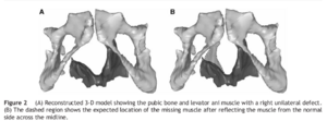
Background/Purpose: Develop a method to quantify the cross-sectional area of the pubic portion of the levator ani muscle, validate the method in women with unilateral muscle defects, and report preliminary findings in those women. METHOD: Multi-planar proton density magnetic resonance images of 12 women with a unilateral defect in the pubic portion of their levator ani were selected from a larger study of levator ani muscle anatomy in women with and without genital prolapse. Three-dimensional bilateral models of the levator ani were reconstructed (using 3-D Slicer, version 2.1b1) and divided into iliococcygeal and pubic portions. Muscle cross-sectional areas were calculated at four equally spaced locations perpendicular to a line drawn from the pubic origin to the visceral insertion using the I-DEAS computer modeling software. RESULTS: The cross-sectional area of the muscle on the side with the defect was smaller than the normal side at all the four locations. The average bilateral difference was up to 81% at location 1 (nearest pubic origin). Almost all of the volume difference (13.7%, P=0.0004) was attributable to a reduction in the pubic portion (24.6%, P<0.0001), not the iliococcygeal portion (P=0.64), of the muscle. CONCLUSIONS: A method was developed to quantify cross-sectional area of the pubic portion of the levator ani perpendicular to the intact muscle direction. Significant bilateral cross-sectional area differences were found between intact and defective muscles in women with a unilateral defect.
Grant Support:
- NICHD R01 HD-38665
A Comparison of Upper Airway Structures in Male and Female Obstructive Sleep Apnoea (OSA) Patients
Publication: 2007 American Thoracic Society International Conference, submitted Oct 2006
Authors: RD McEvoy, MK Ryan, PG Catcheside, M Jakab, A Malhotra, MR Sage, R Valentine, DP White
Institution: Adelaide Institute for Sleep Health, Repatriation General Hospital, SA Australia.
Background/Purpose: For the same BMI female OSA patients tend to have less severe sleep disordered breathing than male OSA patients. This study investigates features of upper airway anatomy that may contribute to this difference. METHOD: Male (n=6) and female (n=9) patients matched for age [mean(SD); men 56.5(5.7), women 55.3(6.2) yrs] and BMI [35.2(3.4), 34.3 (3.0) kg/m2], who had previously been diagnosed with OSA (AHI>30/h), and adherent with CPAP for >3 months were studied. T1 weighted magnetic resonance imaging of the upper airway and surrounding tissues was performed and analyzed (3D-Slicer: www.slicer.org) to determine airway, soft tissue and bony dimensions. RESULTS: The oropharyngeal bony vault volume was greater in males after normalizing for height (1.2(0.1) vs 0.97(0.1), mls/cm, p=0.001). Men had greater neck circumference (45.3(4.3) vs 39.5(4.5) cm, p=0.026) and longer pharyngeal airways (75.2(8.3) vs 64.6(9.5) mm, p=0.042) compared to women. Pharyngeal airway volume and minimum CSA were not different between the genders. In the mid sagittal plane, the tongue CSA was greater in men before and after (36.4(1.4) vs 32.6(3.5) mm²/mm, p=0.01) normalizing for bony vault dimensions. In data collected to date, AHI was not different between males and females (55(12) vs 62(21) events/h). CONCLUSIONS: These preliminary data suggest that male OSA patients have greater oropharyngeal bony vault volume than female patients; a larger tongue (normalized for bony vault dimensions) and a longer pharyngeal airway. These findings may have implications for the male predisposition for OSA.
Grant Support:
- Royal Australian and New Zealand College of Radiologists
Atlas Guided Identification of Brain Structures by Combining 3D Segmentation and SVM Classification
Publication: MICCAI 2006, LNCS 4191, pp. 209–216, 2006. PDF
Authors: Ayelet Akselrod-Ballin, Meirav Galun, Moshe John Gomori, Ronen Basri, and Achi Brandt
Institution: Department of Computer Science and Applied Math, Weizmann Institute of Science, Rehovot, Israel and Dept. of Radiology, Hadassah University Hospital, Jerusalem, Israel
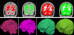
Background/Purpose: This study presents a novel automatic approach for the identification of anatomical brain structures in magnetic resonance images (MRI). The method combines a fast multiscale multi-channel three dimensional (3D) segmentation algorithm providing a rich feature vocab ulary together with a support vector machine (SVM) based classifier. The segmentation produces a full hierarchy of segments, expressed by an irregular pyramid with only linear time complexity. The pyramid provides a rich, adaptive representation of the image, enabling detection of various anatomical structures at different scales. A key aspect of the approach is the thorough set of multiscale measures employed throughout the segmentation process which are also provided at its end for clinical analysis. These features include in particular the prior probability knowl- edge of anatomic structures due to the use of an MRI probabilistic atlas. An SVM classifier is trained based on this set of features to identify the brain structures. We validated the approach using a gold standard real brain MRI data set. Comparison of the results with existing algorithms displays the promise of our approach.
Grant Support:
- Grant #2002/254 Israel Institute of Technology
- IST-2002-506766 European Commission Project
Development of a CAD (Computer Assisted Detection) System to Detect Lung Nodules in CT Scans
Publication: PDF
Authors: Eleonora Tommasi and Giorgio De Nunzio
Institution: Departments of Physics and Material Science, University of Lecce, Italy
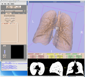
Background/Purpose: We implemented a piece of software to segment lung tissue in CT thoracic images. We gave particular attention to the study of the ilhus pulmonis (region of entrance of bronchi and vessels in the lung parenchima). It is a delicate region in segmentation. We used Slicer to visualize the different steps of the algorithm, checking their correctness. Through a segmentation technique, known as “region growing”, we obtained the delineation of the respiratory apparatus domain in the CT scans. This algorithm is completely three-dimensional. Its output consists in the 3D mask of the respiratory apparatus. We used 3D Slicer to visualize it. Then through an alternative version of region growing, based on a wavefront simulation algorithm, we obtained the segmentation of the external bronchi. We also used the opacity proprieties to put in evidence the internal structures of lungs (such as vessels).
Dynamic Simulation of Joints Using Multi-Scale Modeling
Author: Trent M. Guess
Institution: Mechanical Engineering, University of Missouri-Kansas City
Background/Purpose:
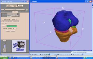
- Develop computationally efficient algorithms that capture tibio-femoral-menisci articulations through neural networks that learn from computationally intensive finite-element tissue models.
- Evaluate tissue interdependencies of the knee during ambulatory activity including the effect of modified menisci properties and/or geometries on tibio-femoral contact pressures.
Grant Support:
- National Science Foundation, Grant Number 506297, under the IMAG program for Multiscale Modeling.
Quasi-isometric Flattening of Curved Surfaces for Medical Imaging
Researchers: Yehoshua Y. Zeevi, Emil Saucan and Eli Appleboim
Institution: Department of Electrical-Engineering, Technion-Israel Institute of Technology, Haifa, Israel.
Description: CT/MRI-scans of human brain-cortex/colon are flattened in a quasi-isometric manner in order to keep angles/length/area as minimally distorted as possible. 3D-surfaces are created out of real data scans, using 3D-slicer .
Grant Support:
- Viterbi Post Doc. fellowship Ollendorf Minerva Center Fund
- Technion V.P. Fund for Promotion of Research at the Technion
3D Multiscale Agent-Based Cancer Model Using MR-Imaging Data
Publication: Zhang L., Athale C.A., Deisboeck T.S. Development of a three-dimensional multiscale agent-based tumor model: Simulating gene-protein interaction profiles, cell phenotypes and multicellular patterns in brain cancer. J Theor Biol. 244(1): 96-107, 2007.
Authors: Thomas S. Deisboeck, Le Zhang, Zhihui Wang
Institution: Complex Biosystems Modeling Laboratory, Massachusetts General Hospital
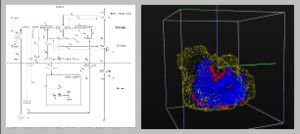
Background/Purpose: Related also to efforts within the Center for the Development of a Virtual Tumor, CViT (http://www.cvit.org), our research here (http://biosystems.mit.edu) focuses on developing multi-scale and multi-resolution (brain) cancer models. Using a hybrid 3D agent-based modeling platform we connect gene-protein interaction maps on the molecular and single cell level (Figure 1, left) up to the volumetric tumor scale of clinical MR-images. For the latter, we plan on using Slicer for segmentation of tumor-affected areas and 3D reconstruction of tumor volume and surface. Amongst the more challenging steps is the translation of these 2D in vivo imaging data onto our in silico lattices. Figure 1 (right) depicts a first step towards this approach where the yellow points depict the 3D surface reconstruction of a T1+Gd MRI while the blue and red points represent virtual tumor cells in attempt to match the real data points.
Grant Support:
- NIH CA 085139
- NIH CA 113004
A Translation Station for Imaging
Publication: RSNA 2006;
Authors: P Mongkolwat, T Lechner, A Kogan, S Talbot, B Johnson, D S Channin
Institution: Imaging Informatics Section, Northwestern University, Chicago, IL 60611, USA
Background/Purpose: Abstract: while providing clinical feedback to the basic sciences. In imaging informatics, this means enabling collaborations between radiologists and computer scientists. Commercial workstations do not allow for experimentation nor do they evolve fast enough. Research imaging packages, while innovative, do not provide for clinical activities or regulatory compliance. We added IHE workflow functionality, hanging protocols and other clinical functions to the SLICER research platform. We leveraged the functionality and methodology of the Insight and Visualization Toolkits, and the DCM4CHE tools. A research mode allows for the evaluation of experimental algorithms against a PACS while protecting patient confidentiality. This combined functionality allows researchers to develop algorithms against clinical cases while at the same allowing clinicians to explore and validate these developments.
Preliminary Study on Digitized Nasal and Temporal Bone Anatomy
Publication: Clin Anat. 2006 Jan; 19(1):32-6. PDF
Authors: Li XP, Han DM, Xia Y, Zhou GH
Institution: Beijing Tongren Hospital Affiliated with Capital University of Medical Sciences, ENT Department, People's Republic of China
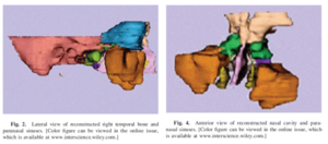
Background/Purpose: The purpose of this study was to explore a feasible method for the reconstruction of the nasal and temporal bone structures of the Chinese virtual human project and provide a more accurate and facilitated way view them three-dimensionally (3D). The 3-D Slicer software was used to reconstruct the anatomic structures of the human nose and temporal bone. Segmentation and extraction of the contours of the ROI (region of interest) in each single slice were conducted and the processed volume data was transferred into the 3-D Slicer. After resegmentation, a set of labeled maps of the ROI were produced. Based on these maps, the 3D surface models of the tissues of interest were constructed. Four groups of paranasal sinuses, nasal septa, middle and inferior turbinates, temporal bones, tympanic cavities, mastoid air cells, sigmoid sinuses, and internal carotid arteries were reconstructed successfully. These models show spatial relationships and orientation between them. The results show that the 3-D Slicer may be used for the 3D visualization of parts of anatomic structures in the nose and temporal bone based on the first Chinese virtual human data, and thus, can facilitate the observation and understanding of the anatomic structures in this area.
Grant Support:
- Beijing Natural Science Fund; Grant number: 7031001
Group-Slicer: a collaborative extension of 3D-Slicer
Publication: J Biomed Inform. 2005 Dec;38(6):431-42. PDF
Authors: Simmross-Wattenberg F, Carranza-Herrezuelo N, Palacios-Camarero C, Casaseca-de-la-Higuera P, Martin-Fernandez MA, Aja-Fernandez S, Ruiz-Alzola J, Westin CF, Alberola-Lopez C
Institution: Laboratorio de Procesado de Imagen, ETSI Telecomunicacion, Universidad de Valladolid, 47011 Valladolid, Spain
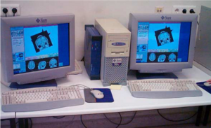
Background/Purpose: In this paper, we describe a first step towards a collaborative extension of the well-known 3D-Slicer; this platform is nowadays used as a standalone tool for both surgical planning and medical intervention. We show how this tool can be easily modified to make it collaborative so that it may constitute an integrated environment for expertise exchange as well as a useful tool for academic purposes.
Grant Support:
- Comision Interministerial de Ciencia y Tecnologıa for Research Grant TIC2001-3808-C02
- NIH P41-RR13218
- CIMIT
- Network of Excellence SIMILAR FP6-507609
Open-configuration MR-guided Microwave Thermocoagulation Therapy for Metastatic Liver Tumors from Breast Cancer
Publication: Breast Cancer. 2005;12(1):26-31.
Authors: Abe H, Kurumi Y,Naka S, Shiomi H, Umeda T, Naitoh H, Endo Y, Hanasawa K, Morikawa S, Tani T.
Institution: Division of General Surgery, Department of Surgery, Shiga University of Medical Science, Seta-Tsukinowa, Otsu, Shiga, 520-2192, Japan.
Background/Purpose: Liver metastases from breast cancer are associated with a poor prognosis, however, local control with microwave thermocoagulation therapy has been used in certain subgroups of these patients in the past decade. In this study, open-configuration magnetic resonance (MR) -guided microwave thermocoagulation therapy was used for metastatic liver tumors from breast cancer, and the efficacy of this treatment was assessed. METHODS: Between June 2000 and April 2004, we used MR-guided microwave thermocoagulation therapy on 11 nodules in 8 patients with metastatic liver tumors from breast cancer. The procedure was carried out under general anesthesia. A 0.5 T open-configuration MR system and a microwave coagulator were used. Near-real-time MR images and real-time temperature images were collected and displayed on the monitor. The MR-compatible thoracoscope was used and combined with MR imaging guidance. Navigation software, a 3D Slicer, was installed and customized. RESULTS: The customized navigation software displayed near-real-time MR images. The percutaneous puncture into the tumors was successful in all cases. No mortality or major complications occurred as a result of the procedures. Five of the 8 patients are alive with new metastatic foci with a mean observation period of 25.9 months. CONCLUSIONS: We developed several devices to allow safe, easy, and accurate MR-guided microwave thermocoagulation therapy of liver tumors. Open-configuration MR-guided microwave thermocoagulation therapy appears to be a feasible method for tumor ablation of metastatic liver tumors from breast cancer.
Virtual Cystoscopy - A Surgical Planning and Guidance Tool
Publication: Arch Ital Urol Androl. 2006 Mar; 78(1):23-24.
Authors: Braticevici B, Onu M, Bengus F.
Institution: Clinical Hospital, Bucharest, Romania.
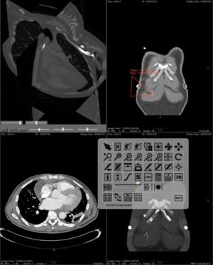
Background/Purpose: Image guided-surgery systems facilitates surgical planning phases of endoscopic procedures. In this paper, we used a software package for 3D surface model generation and vizualization of the urinary bladder, based on magnetic resonance (MR) cross sectional images of the pelvis. The patients group consisted in 6 patients diagnosed with urinary bladder tumour. They were submitted to MRI exam. Twelve consecutive cross sectional images of the pelvis were aquired (TR (repetition time) = 600 msec, TE (echo time) = 19 msec, slice thickness = 6 divided by 7 mm, FOV (field of view) = 36 cm. All these images were transferred to a personal computer running the 3DSlicer software. We obtained, for each patient, a 3D model of the pelvis including the urinary bladder. In This way, the surgical enviroment was simulated and we are able to investigate the bladder by virtual cystoscopy. The virtual endoscopy may be used as a tool in the preoperative training and in surgical planning.
Morphology, constraints, and scaling of frontal sinuses in the hartebeest, Alcelaphus buselaphus (Mammalia: Artiodactlya, Bovidae)
Publication: Journal of Morphology 2007; 268:243-253. PDF
Authors: Farke, A. A.
Institution: Department of Anatomical Sciences, Stony Brook University, N.Y.
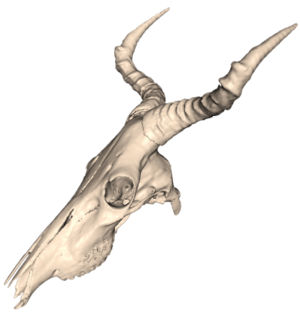
Background/Purpose: ABSTRACT The frontal sinuses of bovid mammals display a great deal of diversity, which has been attributed to both phylogenetic and functional influences. In-depth study of the hartebeest (Alcelaphus buselaphus), a large African antelope, reveals a number of previously undescribed details of frontal sinus morphology. In A. buselaphus, the frontal sinuses conform closely to the shape of the frontal bone, filling nearly the entire element. However, the horncores are never extensively pneumatized, contrasting with the condition seen in many other bovids. This evidence is inconsistent with the hypothesis that sinuses are opportunistic pneumatizing agents, suggesting that phylogenetic factors also play a role in determining sinus size. Both cranial sutures and neurovasculature appear to constrain the growth of sinuses in part. In turn, the sinus also affects the growth of the parietal; apparently this element is not truly pneumatized by the sinus in most cases, but the bone’s shape changes under the influence of the sinus. Furthermore, the sinuses present relatively few struts when compared with the sinuses of some other bovids, such as Ovis. By adapting methods previously developed for measuring structural parameters of trabecular bone, it is possible to quantify certain aspects of sinus morphology. These include the number of bony struts within the sinus, the spacing of these struts, and the size of individual cavities within the sinus. Some differences in the number of struts are evident between subspecies. Similarly, significant differences occur in the relative number of struts between male and female A. buselaphus, which may be related to behavior. The volume of the sinus is strongly correlated with the size of the frontal, but less so with overall cranial size. This finding illustrates the importance of choosing variables carefully when comparing sinus sizes and growth between species.
A ceratopsid dinosaur parietal from New Mexico and its implications for ceratopsid biogeography and systematics
Publication: Journal of Vertebrate Paleontology 2006; 26:1018-1020. PDF
Authors: Farke, A. A., and T. E. Williamson.
Institution: Department of Anatomical Sciences, Stony Brook University, N.Y.
Background/Purpose: Ceratopsid (horned dinosaur) cranial material is relatively common in the Upper Cretaceous Naashoibito Member of the Kirtland Formation, San Juan Basin, New Mexico. However, the specimens are fragmentary, making identification problematic. Previously, it has been suggested that the Naashoibito specimens represent Torosaurus utahensis, a chasmosaurine taxon originally known from the North Horn Formation of Utah. This identification has been used to support a Lancian age for the Naashoibito Member and the Alamo Wash local fauna of the Kirtland Formation (e.g., Lehman, 1981; Lucas et al., 1987). Others (e.g., Lehman, 1990) have synonomized T. utahensis with Torosaurus latus, a taxon otherwise known only from the late Maastrichtian Lance and Hell Creek formations. Recent work has upheld the validity of T. utahensis (Sullivan et al., 2005). However, Sullivan and colleagues (2005) have also argued that no previously described ceratopsid specimens from the Naashoibito Member are identifiable beyond the level of Chasmosaurinae indeterminate and therefore are not useful for assessing biostratigraphic correlations and ceratopsid biogeography. Here we describe a partial chasmosaurine ceratopsid parietal from the Naashoibito Member of the Kirtland Formation. Although other fragmentary parietals have been recovered from this unit (e.g., Lehman, 1981; Lucas et al., 1987), the new specimen exhibits unusual morphology: an epoccipital element positioned along the midline. This feature has previously been reported only in Triceratops, a taxon not known from south of the Denver Basin. The occurrence of such a feature in a ceratopsid from New Mexico has implications for ceratopsian systematics and biogeography.
Grant Support:
- NSF EAR 0207750
