Projects/Diffusion/2007 Project Week Contrasting Tractography Measures/ROI Definitions
|
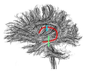
This page describes the definition of the regions of interest for five fiber bundles: the Internal Capsule, the Uncinate Fasciculus, the Fornix, the Arcuate Fasciculus and the Cingulum Bundle.
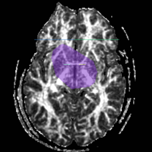
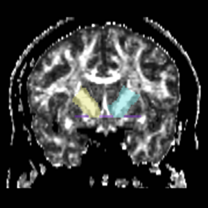
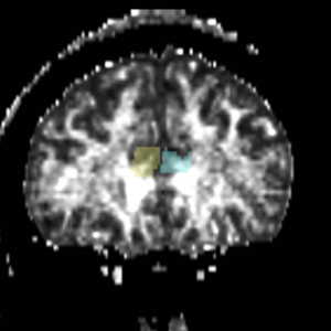
Seed points were defined using a two ROIs approach. Each ROI was drawn on FA and color by orientation maps according to the criteria defined below: ContentsInternal Capsule (IC)ROI1 The inferior boundary of ROI1 was defined by an axial slice containing the anterior commisure (Fig.1). The left and right ROI1s were drawn on a coronal slice containing the anterior commisure, on each side of the midsagittal line (Fig.2). The superior boundary of each ROI1 was defined by the caudate/putamen line |
|
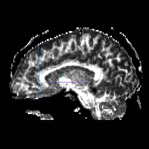
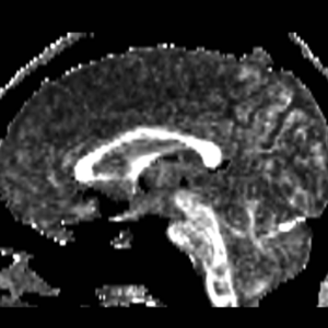
ROI2 The most anterior coronal slice of the corpus collosum was selected using a sagittal view (Fig 3), and the ROI2s were defined by the whole sections of the left and right hemispheres of the brain (Fig 4). |
|
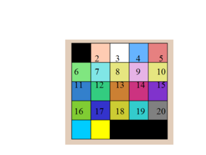
The color coding of the resulting ROIs is as follows:
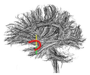
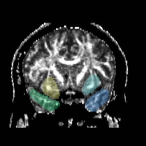
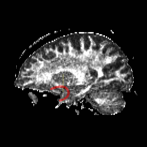
Uncinate Fasciculus (UNC)The most prominant (central) slice of the fornix was identified using a sagittal view (Fig. 5), and the ROI1s and ROI2s were drawn on the coronal slice adjacent to the most anterior point of the fornix (Fig. 6).
|
|
|
|
The color coding of the resulting ROIs is as follows:
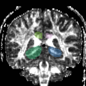
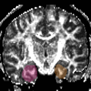
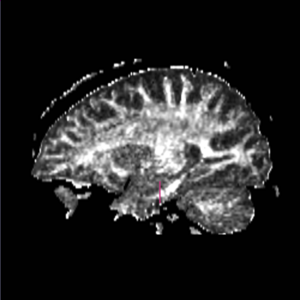
FornixThe ROI1 was drawn on the most anterior coronal slice containing the middle cerebellar peduncles (Fig. 8 & 10). The ROI2 was drawn on a coronal slice posterior to ROI 1, where the middle cerebellar peduncle is clearly connected at bottom of slice (Fig. 9 & 11)
|
|
The color coding of the resulting ROIs is as follows:
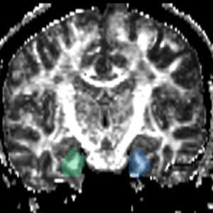
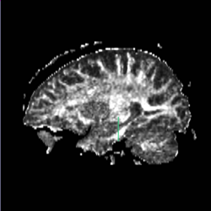
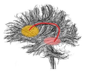
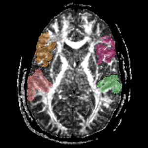
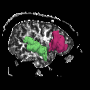
Arcuate FasciculusThese labelmaps ('caseD00XXX-FS-arcuate-final.nhdr') were created using automatic gray matter parcellation in Freesurfer and coregistered in Slicer to corresponding DTI dataset. The labelmaps were dilated in Slicer to increase coverage of gray matter.
|
|
The color coding of the resulting ROIs is as follows:
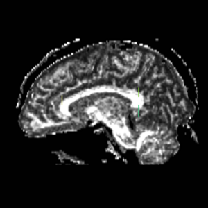
Cingulum BundleROI 1) A coronal plane in the most anterior point of the corpus callosum was selected using the mid-saggital plane (Fig.18), and the left and right ROI1s are drawn on the superior side of the corpus callosum (Fig.15)
|
|
The color coding of the resulting ROIs is as follows:
|
