Slicer Training Volume Rendering
Contents
Volume Rendering
Get It Running
- To get volume rendering to work within 3D Slicer we have to make sure that a MRML node with Volumetric Data exists.
- If not a new set of volume data must be loaded. For this demo we will load the tutorial data found here.
- Download and Uncompress this folder
- In 3D Slicer go to File -> Load Scene
- Select the tutorial.xml
- Now select the VolumeRendering Module.
- Select a valid volume as the Source Volume
The Volume Rendering will be initialized. Have patience....
Parameters
Performance Triggers
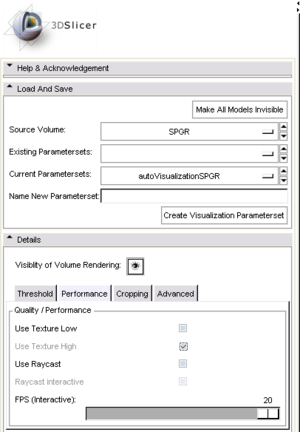
You can adjust the performance of volume rendering by enabling and disabling quality improvements. The Volume Rendering Module has three performance levels
- Low Resolution Hardware Rendering (worst - fastest)
- Full Resolution Hardware Rendering
- Software Rendering (best - slowest)
When all of them are selected slicer uses the low resolution when the camera is moving or nodes in the scene are changing and when nothing is changing anymore the high resolution volume is performed.
The FPS tag lets slicer decide what resolution should be used to produce an interactive experience.
Cropping
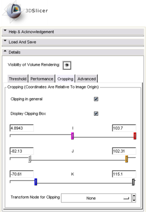
With cropping the volume can be limited to a certain area. This is useful if a surgeon only wants to see a certain part of the volume or cut at an exact plane.
This feature also comes with a integrated 3 dimensional widget to adjust the handlers on the visualization screen.
Try it out!
Advanced Options and the Transfer Function
The advanced tab lets more experienced doctors do the fine tuning on the Transfer Functions.
Transfer Functions are the basic building block of volume rendering. They define the final rendering color for each voxel in the 3D grey scale data. This can be used to give all tissue with the density of bone a solid white color and paint the skin tissue in a translucent pink above it.
More Information
- Slicer3 Volume Rendering Module: Slicer3:Volume_Rendering
- Slicer3 Volume Rendering Module using CUDA: Slicer3:Volume Rendering With Cuda
R. A. Drebin, L. Carpenter, and P. Hanrahan. Volume rendering. pp. 6574, 1988. 3.1 [2] K. Engel, M. Kraus, and T. Ertl. High-quality pre-integrated volume rendering using hardwareaccelerated pixel shading, 2001, citeseer.ist.psu.edu/ engel01highquality.html. 3.1.2
[3] D. T. Gering, A. Nabavi, R. Kikinis, N. Hata, J. ODonnell, W. E. L. Grimson, F. A. Jolesz, P. M. Black, and W. M. W. III. An integrated visualization system for surgical planning and guidance using image fusion and an open MR. J Magn Reson Imag 13(6):967975, 2001. 3.4
[4] H.-R. Ke and R.-C. Chang. Ray-cast volume rendering accelerated by incremental trilinear interpolation and cell templates. The Visual Computer 11:297308, June 2005, http://www.springerlink.com/content/u177877621757257/. 1
[5] J. Kniss, J. Kniss, G. Kindlmann, and C. Hansen. Multidimensional transfer functions for interactive volume rendering. 8(3):270285, 2002. 3.1.3
[6] J. Kniss, J. Kniss, S. Premoze, C. Hansen, P. Shirley, and A. McPherson. A model for volume lighting and modeling. 9(2):150162, 2003. 3.1.4
[7] E. B. Lum, B. Wilson, and K.-L. Ma. High-quality lighting and efficient preintegration for volume rendering. Joint EUROGRAPHICS - IEEE TCVG Symposium
on Visualization, 2004. 3.1.2 [8] K. Martin, W. Schroeder, and B. Lorensen. vtkvolumeraycastcompositefunction from the vtk framework. opensource, freebsd license, http://www.vtk.org. 3.2.1.3 15 Bibliography
[9] N. Max, P. Hanrahan, and R. Crawfis. Area and volume coherence for efficient visualization of 3d scalar functions. SIGGRAPH Comput. Graph. 24(5):2733, 1990. 3.1.2
[10] P. M. Novotny, S. K. Jacobsen, N. V. Vasilyev, D. T. Kettler, I. S. Salgo, P. E. Dupont, P. J. D. Nido, and R. D. Howe. 3d ultrasound in robotic surgery: performance evaluation with stereo displays. Int J Med Robot 2(3):279285, Sep 2006, http://dx.doi.org/10.1002/rcs.102. 4
[11] P. M. Novotny, D. T. Kettler, P. Jordan, P. E. Dupont, P. J. del Nido, and R. D. Howe. Stereo display of 3d ultrasound images for surgical robot guidance. Conf Proc IEEE Eng Med Biol Soc 1:15091512, 2006, http://dx.doi.org/ 10.1109/IEMBS.2006.259486. 4
[12] P. M. Novotny, J. A. Stoll, N. V. Vasilyev, P. J. del Nido, P. E. Dupont, T. E. Zickler, and R. D. Howe. Gpu based real-time instrument tracking with three-dimensional ultrasound. Med Image Anal 11(5):458464, Oct 2007, http: //dx.doi.org/10.1016/j.media.2007.06.009. 4
[13] NVidia. NVIDIA CUDA Compute Unified Device Architecture - Programming Guide. NVidia Corporation, 1.1 edition, Nov 2007, http://www.nvidia.com/ cuda. 1 16


