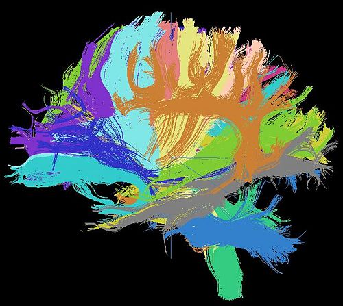
2009 Winter Project Week Tractography Segmentation
 Return to Project Week Main Page |
Key Investigators
- Xiaogang Wang, MIT
- Carl-Fredrik Westin, BWH
Objective
We are developing a nonparametric Bayesian approach to cluster white matter fiber tracts to bundles. These bundles correspond to white matter anatomy. This approach is used to cluster fibers across multiple subjects and compare their anatomical structures. The number of clusters is learnt from data instead of being manually specified.
Approach
Our approach models bundles as multinomial distributions over voxels and orientations. Under a hierarchical Bayesian model, Dirichlet process is used as a prior distribution to learn the number of clusters. Gibbs sampling is used for inference.
Our plan is to integrate this approach into 3D-slicer, use 3D-slicer to do experimental evaluation (including registering multiple subjects, generating fibers and clustering fibers), and compare with the approach proposed by [1],2], which is based on spectral clustering and the mean of closest distances.
Progress
We have the code of our new approach written in C++ and matlab. In this project week, we plan to integrate our code into 3D-slicer. We also have a numpy-slicer module developed by Demian Wasserman that implements [2]. We plan to use this code as a template when porting our new method to numpy/slicer.
References
[1] Brun A, Knutsson H, Park HJ, Shenton ME, Westin CF. Clustering fiber tracts using normalized cuts. In Seventh International Conference on Medical Image Computing and Computer-Assisted Intervention (MICCAI'04), Lecture Notes in Computer Science. Rennes - Saint Malo, France, 2004;368-375.
[2] O'Donnell LJ, Westin CF. Automatic tractography segmentation using a high-dimensional white matter atlas. IEEE Transactions on Medical Imaging 2007;26(11):1562-1575.
