OrientationTestData
From NAMIC Wiki
Home < OrientationTestData
Bill Lorensen collected a set of MR data sets a various standard orientations of a cardiac phantom. Because correct orientation information in image file headers is crucial in medical imaging, these images are being made available as a reference for the community.
The data includes the following scan orders:
- A->P, P->A, L->R, R->L, I->S, S->I
(L = left, R = right, A = anterior, P = Posterior, I = inferior, S = superior).
The data is available for download and testing:
Notes:
- the scans are all of the same phantom, but have different fields of view, so even if you have the scan orientations correct, the images won't line up pixel for pixel (the rigid registration matrices allow direct comparison).
- the cardiac phantom contains some symmetry, so be careful when making comparisons
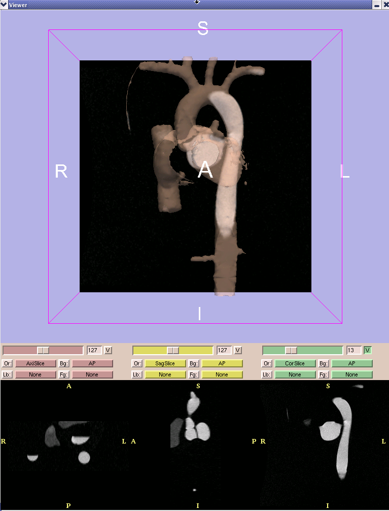
- the screenshot from slicer below shows the correct orientation for the images (see the label letters for reference). A semi-transparent rendering of a 3D model of the phantom is shown for reference. The model is included with the scene files above.
- The Slicer scenes were made by reading the dicom images with the ITK-based reader. The .nii images were made by saving with the ITK-based NIfTI writer. The transformations were calculated using the interactive ITK-based rigid registration tools in Slicer.
Additional formats for these images, generated for example by different processing tools, would be welcome. Other images that could be used to help debug/verify orientation issues in headers would be welcome.
