Mbirn: Gradient non-linearity distortion correction
- Short Description
The goal of this software is to provide a 3D correction of image distortions in MRI data due to non-linearity of the magnetic fields from the imaging gradient coils.
Keywords: MRI distortions, gradient non-linearities, multi-site studies, longitudinal studies

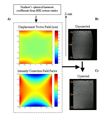
Figure: Schematic representation of the distortion correction process. The spherical harmonic coefficients specific to the MR system are used to generate 3D x,y,z displacement fields (here we show the magnitude) and intensity correction fields arising from voxel distortions (Figure 1A). The correction fields correspond to a 300mm x 300mm coronal slice through and centered on the scanner isocenter. The distortions on the uncorrected phantom image appear as diameter variations along the z-axis (Figure 1B). These geometric distortions are significantly reduced in the corrected image (Figure 1C).
- Longer Overview
- Longitudinal and multi-site clinical studies create the imperative to characterize and correct technological sources of variance that limit image reproducibility in high-resolution structural MRI studies, thus facilitating precise, quantitative, platform-independent, multi-site evaluation. An important task in this effort is to correct for site-specific image distortions in order to allow accurate cross-site comparisons of quantitative morphometry results. While in principle distortions from gradient non-linearities may be addressable using manufacturer-supplied software, the currently available correction algorithms work only in two-dimensions (2D) providing an incomplete solution to the problem
- The software distributed provides a 3D distortion correction based on knowledge of the spherical harmonic coefficients from the imaging gradients.
- Outline of steps involved to complete gradient unwarping and contents of this distribution:
- Obtain from your local Siemens or GE vendor the spherical harmonics expansion that represents the gradient coils used in the MRI system you want to correct. This is a text file with proprietory information.
- We provide a pearl script (gradient_unwarp_converter.pl) that reads in the spherical harmonics text file and writes out the coefficients in a standard format (for example, avanto.coef). This script needs to be run only once per gradient system.
- ) We provide a script (create_displacement_table) that reads in the standard formatted coefficients (e.g., avanto.coef) of your system and writes out a file that has the tables for displacements along the x,y,z axes and also the intensity correction table due to voxel size distortion (e.g., avanto.gwv). This script needs to be run only once per gradient system.
- We provide a script (grad_unwarp) that reads in the correction tables of your system and the dicom image volume you want to correct and writes out the distortion corrected dicom volume. For more details on other image formats see 'Use instructions' below.
- We also provide: - Sample phantom data, both raw and distortion corrected, for different platforms and gradient systems (Siemens Sonata, GE CRM) - An image format conversion tool (mri_convert) - A matlab file viewer for 'mgh' formatted image volumes
- User Manual and Installation Instructions
- The distribution includes a README text file with use and installation instructions
- Registration for Download
- References
- Jorge Jovicich, Silvester Czanner; Douglas Greve; Elizabeth Haley; Andre van der Kouwe; Randy Gollub; David Kennedy; Franz Schmitt; Gregory Brown; James MacFall; Bruce Fischl; Anders Dale. Reliability in Multi-Site Structural MRI Studies: Effects of Gradient Non-linearity Correction on Phantom and Human Data, NeuroImage (in press)
- J. Jovicich, S. Czanner, D. Greve, J. Pacheco, E. Busa, A. van der Kouwe, Morphometry BIRN, B. Fischl. Test-retest reproducibility assessments for longitudinal studies: quantifying MRI system upgrade effects. In: International Society of Magnetic Resonance in Medicine; Miami, FL.;2005.
- Technical Contacts
- Silvester Czanner (czanner@nmr.mgh.harvard.edu)
- Jorge Jovicich (jovicich@nmr.mgh.harvard.edu)
- Acknowledgements
The authors thank: a) our human phantoms, b) Steve Pieper and Charles Guttmann (Brigham Women's Hospital) for coordinating the phantom scans at BWH, c) Gabriela Czanner (Massachusetts General Hospital) for her support on statistical analyses and d) Gary Glover (Stanford University), Larry Frank (University of California, San Diego) and Jason Polzin (GE Healthcare Technologies), Eva Eberlein (Siemens Medical Solutions, Erlangen, Germany), for their support on making available the gradient's spherical harmonics information. This research was supported by a grant (#U24 RR021382) to the Morphometry Biomedical Informatics Research Network (BIRN, http://www.nbirn.net), that is funded by the National Center for Research Resources (NCRR) at the National Institutes of Health (NIH).