2011 Winter Project Week:Osteomark
Key Investigators =
- Nobuhiko Hata, Laurent Chauvin
Objective
Develop and validate Osteoplan module in 3D Slicer as part of SBIR Phase II (PI: Magill). 'Abstract from the grant. DESCRIPTION (provided by applicant): Having successfully completed a Phase I program, we propose to continue the development and demonstration of osteomark, a tool for implementing computer-based plans for orthognathic surgery. Physical Sciences Inc., is leading the development of the product with support from our clinical partners in the Dept of Oral and Maxillofacial Surgery at the Massachusetts General Hospital and image-guided surgery experts in the Surgical Planning Laboratory at Brigham and Women's Hospital. The device can be used in both endoscopic and open procedures to transfer treatment plans from computer-based 3D surgical planning tools onto the bone. The system will provide for intraoperative registration of the image to the patient, with no requirement for fiducial markers in the pre-operative images and no fixation frame attached to the head. The surgeon will mark the bone for ostotomies, screw holes, and alignment, and then complete the procedure using instruments without any tracking, using the marks to guide the modification of bone. The proposed device will be less expensive and less complex than a system that tracks the motion of multiple surgical instruments as procedures are performed. osteomark is being developed to interact with the open-source medical imaging software Slicer/Osteoplan, though integration with an FDA-approved commercial program is planned for late in Phase II. In Phase I, we demonstrated the ability to transfer treatment plans to the mandible in endoscopic surgery on a pig cadaver. In Phase II, we will refine the system components and algorithms, carry out several operations on cadavers, and then perform tests on live minipigs. PUBLIC HEALTH RELEVANCE: Minimally-invasive surgery is becoming more widely used in reconstruction of bones in the face because it results in faster recovery times, less pain and discomfort, and less scarring than open procedures. Another recent advancement has been the use of computer graphics tools to plan complex reconstruction operations. We are proposing to develop a device that will enable surgeons to copy computer treatment plans to the bone even through a very small incision, allowing surgeons to perform more sophisticated procedures with less difficulty for the patient and improved outcomes.
Approach, Plan
- Understand the work in Slicer 2
- Discuss requirement of the Ostomark
- Investigate functions, modules in 3D Slicer meeting the requirements
- Design workflow of the OsteoPlan
- Code!
Progress
http://www.na-mic.org/Wiki/index.php/OsteoPlan
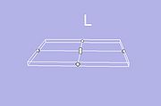
- Cutter has been created (with vtkBoxWidget)
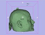
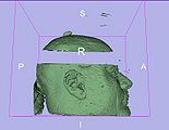
- The clipping model with the cutter has been made (with vtkClipPolyData)
- Finally, we obtain a new model with a part removed (place of the cutter) depending on the thickness of the cutter. The two parts created of the model after clipping couldn't be move separatly
- Improvements could be done
- Fix 2 bugs on vtkBoxWidget (size of handles when resizing and surface rendering)
- Try to improve clipping speed
- Add a option for the thickness of the cutter (need to be discused to see if necessary)


