DBP3:Utah:RegCases
From NAMIC Wiki
Home < DBP3:Utah:RegCases
Back to Utah AFib DBP
Background
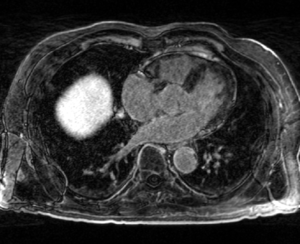
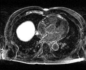
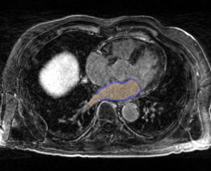
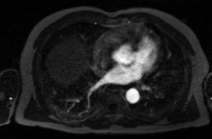
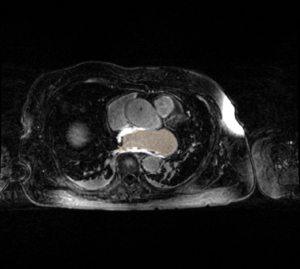
- The CARMA Center uses late gadolinium enhanced MRI (LGE-MRI) images to evaluate new patients, predict procedural success, and evaluate therapeutic outcomes. The MRI images for each patient are further accompanied by MR angiographic images (MRA) and manual segmentations of relevant structures. These images are acquired longitudinally over the course of a patient's evaluation, treatment, and follow-up (i.e. months or years). Registration is often necessary to compare images from different time points in a patient's treatment, across patient cohorts at the same stage of disease progression or treatment, or different image types.
| Pre-ablation LGE-MRI Image | Post-ablation LGE-MRI Image | Segmentation of LGE-MRI Image |
|---|---|---|
| MRA Image | Immediately Post-ablation (IPA) LGE-MRI Image |
|---|---|
Registration Case Types
Pre-ablation LGE to 3 months (or more) post-ablation LGE
- Examine the location of ablation-induced scar formation relative to fibrosis
- Utah score staging of patients [1]
Pre-ablation LGE to Immediately post-ablation(IPA) LGE
- Can compare acute ablation-induced changes to the pre-ablation tissue
Immediately post-ablation to 3 months (or more) post-ablation LGE
- Dark regions on IPA LGE scans result in stable scar formation at 3 or more months post-ablation -> [2]
- Look for gaps in the ablation lesion sets - Ravi's project
Post-ablation LGE to Post-ablation LGE
- Comparing the outcome of repeated ablation procedures to the
Pre-ablation LGE to Pre-ablation LGE
- Comparison across patients prior to ablation
LGE to Dark-blood MRI
- Dark blood images can be used to detect early lesion formation
- Can compare with lesion formation seen later on LGE images
LGE to MRA
- The boundaries of the LA in the MRA mirror the endocardial surface in the LGE scans
- There is the possibility for thresholding the MRA and then registering the segmented region to the LGE
LGE to Electroanatomic map (Carto)
- Compare the low voltage regions of Carto maps to the LGE scans
LGE to CT
- Align LGE and CT for those patients that have both
- Maybe helpful for shape analysis studies
Pre-ablation LGE/Endo to Post-ablation LGE/Endo
- Combined the LGE images and manual segmentations can be used to improve the quality of registration
- Yi Gao previously developed a Slicer module to register pre- and post-ablation images driven by the LGE images/segmentations -> AFib Registration
Implementation
- We need to define a suitable set of registration parameters for each of the above cases
- We will develop a Slicer extension module with the pre-defined registration parameters for each of the above scenarios
