Projects:DTIClustering
Project Description
Collaborators: Lauren O'Donnell (MIT), Carl-Fredrik Westin (BWH), Karthik Krishnan (Kitware), Martha Shenton (Harvard).
In the past, we have demonstrated ways to characterize the strength of connectivity between selected regions in the brain based on several alternative ways to integrate local diffusion tensor measurements into a global field that provided connection strength estimates for distant points. Our current work aims to provide structural description of the white matter as partitioned into coherent fiber bundles and clusters.
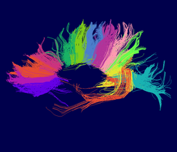
We are developing tools in the 3D Slicer for automatic clustering of tractographic paths through diffusion tensor MRI (DTI) data. By grouping tractographic paths based on shape and location, the white matter architecture may be more clearly visualized, and interesting properties of the clusters (such as for example FA or Westin's linear measure) may be quantified.
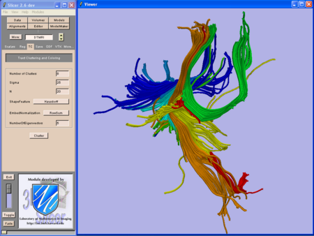
The above image shows the slicer clustering interface and example clusters.
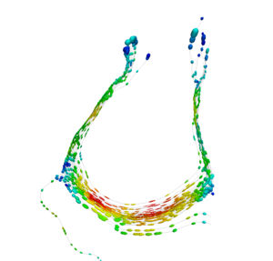
This image shows the same paths as the bright green cluster in the previous image (part of the corpus callosum). Here randomly sampled tensors are displayed along the paths and colored with Westin's linear measure.
Clustering Implementation
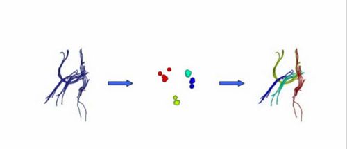
Our implementation uses spectral clustering, a method for grouping data using eigenvectors of a data affinity matrix. This image gives an overview of the method.
On the left example input tractographic paths are shown (these were created by manually seeding in the 3D Slicer). The center image shows an embedding of the tracts as points in 2D, where the distance between points is related to their shape similarity. This embedding was calculated as an intermediate step during spectral clustering. The image on the right shows the final output in the 3D Slicer, where tractographic paths are colored by cluster membership.
Recent Update
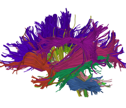
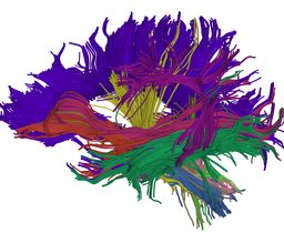
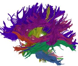
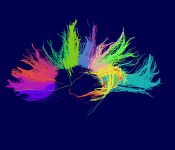
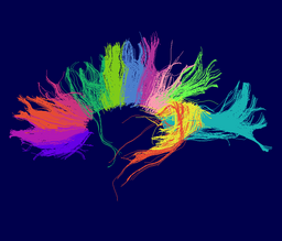
Atlas creation and automatic labeling has been performed in high-quality DTI datasets from Susumu Mori. Images showing example segmentation results are below. Work is underway to apply this atlas to segment additional datasets to define regions of interest that may be used in the study of schizophrenia.
Example Results:
Selected anatomical regions, automatically labeled using the cluster atlas in 3 subjects.
Subdivisions of the corpus callosum, labeled using the cluster atlas in 3 subjects.
Publications
Lauren O'Donnell and Carl-Fredrik Westin. High-Dimensional White Matter Atlas Generation and Group Analysis. MICCAI, 243-251, 2006 (pdf) Lauren O'Donnell. Cerebral White Matter Analysis Using Diffusion Imaging. Harvard-MIT Division of Health Sciences and Technology PhD Thesis, May 2006. (pdf)
Lauren O'Donnell, Carl-Fredrik Westin. A High-Dimensional Fiber Tract Atlas. International Society of Magnetic Resonance in Medicine (ISMRM) 2006. [/Wiki/images/2/2b/Odonnell_WMTractAtlas_ismrm2006-1.pdf (pdf)]
Lauren O'Donnell, Marek Kubicki, Martha E. Shenton, Mark E. Dreusicke, W. Eric L. Grimson, Carl-Fredrik Westin. A Method for Clustering White Matter Fiber Tracts. American Journal of Neuroradiology (AJNR) 27(5):1032-1036, 2006. [/Wiki/images/6/6d/ODonnell_AJNR2006-1.pdf (pdf)]
Lauren O'Donnell, Carl-Fredrik Westin White Matter Tract Clustering and Correspondence in Populations Conference on Medical Image Computing and Computer-Assisted Intervention (MICCAI), 140-147, 2005
Lauren O'Donnell, W. Eric L. Grimson, Carl-Fredrik Westin. Interface Detection in Diffusion Tensor MRI. MICCAI, 360-367, 2004.
Lauren O'Donnell, Steven Haker, and Carl-Fredrik Westin. New Approaches to Estimation of White Matter Connectivity in Diffusion Tensor MRI: Elliptic PDEs and Geodesics in a Tensor-Warped Space. MICCAI, 459-466, 2002.
Lauren O'Donnell, Carl-Fredrik Westin White Matter Tract Clustering and Correspondence in Populations MICCAI, 140-147, 2005. (pdf)
Software
The software employed in this process includes:
- Slicer DTMRI module VTK classes for tractography.
- Slicer VTK class to compute tract path affinity matrix.
- This matrix contains results of shape comparisons between tract paths.
- Several methods are available: endpoint distance, Hausdorff distance, and mean/covariance distance.
- New ITK class (itkSpectralClustering) clusters data based on an affinity matrix.
- Generally applicable for clustering problems because input is just this matrix.
- New and improved ITK Statistics framework which allows our embedding vectors (such as those in the middle image above) to have variable length and therefore employ more shape information when it is present. Thank you to Karthik Krishnan for reworking the ITK Statistics classes.
All of the code is part of NA-MIC. The VTK classes are in the 3D Slicer DTMRI module, while the ITK class is currently located in the NA-MIC Sandbox and will be included in ITK in the future.
Additional matlab code is used for multiple subject clustering and atlas labeling.
