2017 Winter Project Week/MeningiomaSegmentation

From NAMIC Wiki
Revision as of 21:20, 13 January 2017 by Jakubk (talk | contribs)
(diff) ← Older revision | Latest revision (diff) | Newer revision → (diff)
Jump to: navigation, search
Home < 2017 Winter Project Week < MeningiomaSegmentation

Key Investigators

  • Jakub Kaczmarzyk, MIT
  • Satrajit Ghosh, MIT
  • Omar Arnaout, Brigham and Women's Hospital

Project Description

Objective Approach and Plan Progress and Next Steps
  • Segment meningiomas in structural MR images.
  • Assess state of brain after surgical removal of meningioma.
  • Evaluate performance of existing segmentation methods.

Progress

  • Segmented with ANTs and FSL.
  • Learned about Slicer segmentation tools, and segmented semi-automatically.
  • Put in contact with people who have segmented meningiomas.

Next steps

  • Continue learning about what has been done in the past.
  • Improve brain-extraction.
  • Continue testing existing segmentation methods.
  • Try Slicer in a Nipype workflow.
  • Apply feature engineering techniques, like manifold learning.
  • Get more data, and potentially train a neural network.


Examples

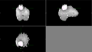
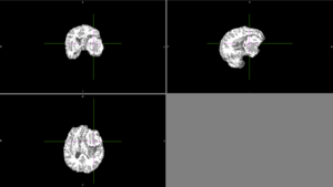
Sometimes, ANTs failed to remove parts of the skull close to the tumor or wrongly removed part of the brain.
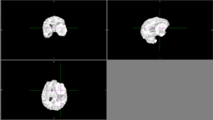
Other times, ANTs extracted the brain well.
Automatic segmentation with ANTs usually could not distinguish the tumor from the rest of the brain.
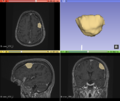
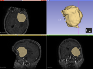
Semi-automatic segmentation with Slicer was relatively successful. Sometimes the segmentation would bleed outside of the tumor into voxels with similar intensities.

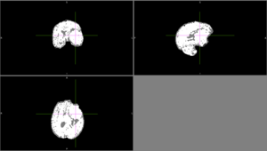
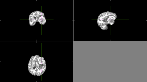
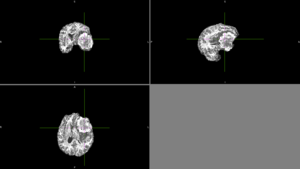
We tried FSL's FAST with different numbers of classes. None of these methods could identify the entire tumor mass as one type of tissue in this scan.

FAST brain segmentation with 3 classes
FAST brain segmentation with 4 classes
FAST brain segmentation with 5 classes


Background and References

MR images of meningiomas that will be used in this project are available at OpenNeu.ro.