Difference between revisions of "2009 Summer Project Week Adaptive RT"
From NAMIC Wiki
Ivan.kolesov (talk | contribs) |
Ivan.kolesov (talk | contribs) |
||
| (10 intermediate revisions by the same user not shown) | |||
| Line 1: | Line 1: | ||
__NOTOC__ | __NOTOC__ | ||
| − | + | ||
| − | Image:PW2009-v3.png|[[2009_Summer_Project_Week#Projects|Projects List]] | + | |
| − | + | ||
| + | |||
| + | {| | ||
| + | |[[Image:PW2009-v3.png|thumb|320px|Return to [[2009_Summer_Project_Week#Projects|Projects List]] ]] | ||
| + | |[[Image:MandibleInitCut.JPG|thumb|320px|Initial Curve]] | ||
| + | |[[Image:MandibleSegCut.JPG|thumb|320px|Final Segmentation]] | ||
| + | |} | ||
| + | |||
==Key Investigators== | ==Key Investigators== | ||
| Line 35: | Line 42: | ||
<h3>Progress</h3> | <h3>Progress</h3> | ||
| − | + | *Segmentation results are shown above. | |
| + | *We have three potential approaches to segmenting the brain stem to experiment with. | ||
| + | *Created a complete data set for one patient including label maps for structures of interest. | ||
Latest revision as of 20:45, 25 June 2009
Home < 2009 Summer Project Week Adaptive RT
 Return to Projects List |
Key Investigators
- GaTech: Ivan Kolesov, Vandana Mohan, and Allen Tannenbaum
- MGH: Gregory Sharp
Objective
We are developing strategies to perform segmentation of a number of structures in the Head, Neck, and Thorax. Once segmentation is available, the goal is to register patient scans to account for anatomical changes between visits.
Approach, Plan
- Get an accurate segmentation before beginning work on registration
- First structure of interest is the mandible, by the end of the project week, obtain its segmentation
- Come up with a strategy for segmenting the brain stem
- Create an outline for further progress after the project week
Progress
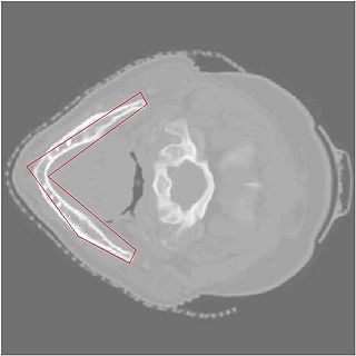
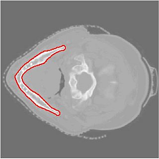
- Segmentation results are shown above.
- We have three potential approaches to segmenting the brain stem to experiment with.
- Created a complete data set for one patient including label maps for structures of interest.

