2009 Summer Project Week MRSI-Module
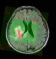
Color-coded MRSI in the localization and grading of brain tumors

Fitting metabolite models to the MRS signal of tumorous brain tissue (top: baseline removal; bottom: resonance line model fits)


Key Investigators
- MIT: Bjoern Menze, Polina Golland
- Iowa: Jeff Yager, Vince Magnotta
Objective
Magnetic resonance spectroscopic imaging (MRSI) is a non-invasive diagnostic method used to determine the relative abundance of specific metabolites at arbitrary locations in vivo. Certain diseases -- such as tumors in brain, breast and prostate -- can be can be associated with characteristic changes in the metabolic level.
The objective of the current project is to develop a module proving the means for the processing and visualization of MRSI -- and thus for a joint analysis of magnetic resonance spectroscopic images together with other imaging modalities -- in Slicer.
Approach, Plan
Spectral fitting routines have been implemented, using a HSVD filter for water peak removal and baseline estimation, and a constrained non-linear least squares optimization for the fit of the resonance line models. MRSI post-processing routines for partial volume correction are available from the BrainCSI toolbox.
Our plan for the project week is to first integrate the spectral fitting routines into a Slicer module and to validate this module using MRSI phantom data. We then want to work towards interfacing signal processing module and post-processing tools.