Difference between revisions of "2010 Summer Project Week Quantification Of Lesion Diffusion Disruption"
From NAMIC Wiki
(Initial page) |
(Added FA distributions image) |
||
| (3 intermediate revisions by the same user not shown) | |||
| Line 2: | Line 2: | ||
<gallery> | <gallery> | ||
Image:PW-MIT2010.png|[[2010_Summer_Project_Week#Projects|Projects List]] | Image:PW-MIT2010.png|[[2010_Summer_Project_Week#Projects|Projects List]] | ||
| − | Image: | + | Image:TractsLesionZoomOverlay.png|Tracts interrupted by lesions with FA values. |
| − | Image: | + | Image:Fa_dists.png|FA distributions for voxels near or inside lesions. |
</gallery> | </gallery> | ||
| Line 27: | Line 27: | ||
<h3>Approach, Plan</h3> | <h3>Approach, Plan</h3> | ||
| − | Our initial approach for quantifying diffusion interruption is to compile statistics on the distributions of FA at varying distances from lesion tissue. Assuming there are significant differences more advanced methods | + | Our initial approach for quantifying diffusion interruption is to compile statistics on the distributions of FA at varying distances from lesion tissue. Assuming there are significant differences more advanced methods will be investigated. |
</div> | </div> | ||
| Line 34: | Line 34: | ||
<h3>Progress</h3> | <h3>Progress</h3> | ||
| − | Using diffusion tools in slicer FA images were created in the same space as the lesion images. ITK code for summarizing statistics is | + | Using diffusion tools in slicer FA images were created in the same space as the lesion images. ITK code for summarizing statistics is complete and analysis of all lupus subjects is under way. |
</div> | </div> | ||
Latest revision as of 14:05, 25 June 2010
Home < 2010 Summer Project Week Quantification Of Lesion Diffusion Disruption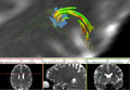
Tracts interrupted by lesions with FA values.
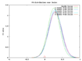
FA distributions for voxels near or inside lesions.


Key Investigators
- ABMIG: H. J. Bockholt
- MRN: Mark Scully
Objective
Quantify the effect lesions have on diffusion.
Approach, Plan
Our initial approach for quantifying diffusion interruption is to compile statistics on the distributions of FA at varying distances from lesion tissue. Assuming there are significant differences more advanced methods will be investigated.
Progress
Using diffusion tools in slicer FA images were created in the same space as the lesion images. ITK code for summarizing statistics is complete and analysis of all lupus subjects is under way.
Delivery Mechanism
This work will be delivered to the NA-MIC Kit as a Slicer3 commandline extension.