2011 Winter Project Week:TubeTK VascularImageSegmentationAndAnalysis
From NAMIC Wiki
Home < 2011 Winter Project Week:TubeTK VascularImageSegmentationAndAnalysis

Tortuosity for benign/malignant (Image from Dr. Bullitt, UNC)
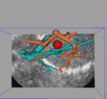
Ultrasound to vessel registration
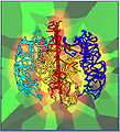
Spatial graphs of vasculature capture inter-population variations


Key Investigators
- Kitware: Stephen Aylward, Danielle Pace
- SPL: Steve Pieper
- Luca Antiga, Daniel Haehn
Objective
TubeTK is a new open-source toolkit that hosts algorithms for applications involving images of tubes.
Two driving applications:
- Surgical guidance: registering pre-operative vascular models with intra-operative images (e.g., ultrasound)
- Characterizing vascular patters: using graph theory to distinguish clinical populations based on vascular patterns (e.g., benign -vs- malignant tumors via tortuosity)
History
- June 2001, UNC released the patent on vessel extraction method from [Aylward, Bullitt 1996...]
- TubeTK released under Apache 2.0 license: includes rights to patents
Approach, Plan
- Python module in Slicer 4 for centerline and radius estimation of vasculature in brain MRA
- Workflow: brain envelop segmentation, seeding, extraction
- Integration with VMTK
Progress
- Skype meeting with VMTK team to learn design pattern to follow
- Extended TubeTK to include LDA methods for multi-echo MR segmentation
- SWAN (susceptibility weighted angiography), T1, T2 data from U of Mississippi
Delivery Mechanism
This work will be delivered to the NA-MIC Kit as follows:
- All software written during the project week will be contributed to TubeTK, and algorithms will be incorporated into 3D Slicer as CLI applications.