Difference between revisions of "2012 Summer Project Week:Interactive Needle Segmentation"
(Created page with 'coming') |
|||
| (10 intermediate revisions by 3 users not shown) | |||
| Line 1: | Line 1: | ||
| − | + | __NOTOC__ | |
| + | <gallery> | ||
| + | |||
| + | |||
| + | Image:PW-MIT2012.png|[[2012_Summer_Project_Week#Projects|Projects List]] | ||
| + | Image:gyn-needle-segmentation-1.png | Screenshot of Manual Needle Segmentation (20 minutes) | ||
| + | Image:Needle-TractographyFiducialSeeding.png | Screenshot of Single Needle Modeled With Tractography Fiducial Seeding Module | ||
| + | Image:Needle-TractographyFiducialSeeding2.png | Screenshot of Multiple Needles Modeled With Tractography Fiducial Seeding Module (Hessian Image Visible) | ||
| + | </gallery> | ||
| + | |||
| + | ==Key Investigators== | ||
| + | *Rutgers: Nabgha Farhat | ||
| + | *BWH: Yi Gao, Xiaojun Chen, Neha Agrawal, Nabgha Farhat, Jan Egger, Tina Kapur, Akila Viswanathan | ||
| + | *Isomics: Steve Pieper | ||
| + | |||
| + | <div style="margin: 20px;"> | ||
| + | <div style="width: 27%; float: left; padding-right: 3%;"> | ||
| + | |||
| + | <h3>Objective</h3> | ||
| + | The goal of this project is to achieve fast (< 2 minute) interactive segmentation of 5-10 needles from MRI images using Slicer. | ||
| + | |||
| + | |||
| + | </div> | ||
| + | |||
| + | <div style="width: 27%; float: left; padding-right: 3%;"> | ||
| + | |||
| + | <h3>Approach, Plan</h3> | ||
| + | |||
| + | We will explore different segmentation/editing tools in Slicer to determine which ones can most efficiently segment 2mm needles (10-15 cm long), from MRI scans in which voxels are about 2mm in each dimension. (These needles are not straight lines, and are often placed within a few mm of each other.) | ||
| + | |||
| + | </div> | ||
| + | |||
| + | <div style="width: 40%; float: left;"> | ||
| + | |||
| + | <h3>Progress</h3> | ||
| + | |||
| + | Needles were effectively modeled from pelvic MR scans using the Tractography Fiducial Seeding module: | ||
| + | *Hessian images were generated from the MR scans | ||
| + | *Fiducials were placed in regions known to be part of a needle | ||
| + | *The Tractography Fiducial Seeding module was used to generate “tracts” that pass through the fiducial – in this case, the needles | ||
| + | Refinements need to be made to optimize the modeling for brachytherapy needles rather than fiber bundles: | ||
| + | *A sense of “momentum” is needed, so that needles do not abruptly end or change direction in signal voids | ||
| + | *Eventually, we would like the whole workflow to take place in Slicer, including the generation of the hessian image | ||
| + | |||
| + | |||
| + | |||
| + | </div> | ||
| + | </div> | ||
| + | |||
| + | <div style="width: 97%; float: left;"> | ||
| + | |||
| + | ==Delivery Mechanism== | ||
| + | |||
| + | N/A | ||
| + | |||
| + | ==References== | ||
| + | |||
| + | |||
| + | </div> | ||
Latest revision as of 02:20, 26 June 2012
Home < 2012 Summer Project Week:Interactive Needle Segmentation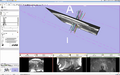
Screenshot of Manual Needle Segmentation (20 minutes)
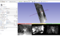
Screenshot of Single Needle Modeled With Tractography Fiducial Seeding Module
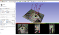
Screenshot of Multiple Needles Modeled With Tractography Fiducial Seeding Module (Hessian Image Visible)


Key Investigators
- Rutgers: Nabgha Farhat
- BWH: Yi Gao, Xiaojun Chen, Neha Agrawal, Nabgha Farhat, Jan Egger, Tina Kapur, Akila Viswanathan
- Isomics: Steve Pieper
Objective
The goal of this project is to achieve fast (< 2 minute) interactive segmentation of 5-10 needles from MRI images using Slicer.
Approach, Plan
We will explore different segmentation/editing tools in Slicer to determine which ones can most efficiently segment 2mm needles (10-15 cm long), from MRI scans in which voxels are about 2mm in each dimension. (These needles are not straight lines, and are often placed within a few mm of each other.)
Progress
Needles were effectively modeled from pelvic MR scans using the Tractography Fiducial Seeding module:
- Hessian images were generated from the MR scans
- Fiducials were placed in regions known to be part of a needle
- The Tractography Fiducial Seeding module was used to generate “tracts” that pass through the fiducial – in this case, the needles
Refinements need to be made to optimize the modeling for brachytherapy needles rather than fiber bundles:
- A sense of “momentum” is needed, so that needles do not abruptly end or change direction in signal voids
- Eventually, we would like the whole workflow to take place in Slicer, including the generation of the hessian image
Delivery Mechanism
N/A