Difference between revisions of "2012 Summer Project Week:QuantitativePETImageAnalysisModule"
(Created page with '__NOTOC__ <gallery> Image:PW-MIT2012.png|Projects List Image:genuFAp.jpg|Scatter plot of the original FA data through the genu of the corpus…') |
|||
| (8 intermediate revisions by the same user not shown) | |||
| Line 2: | Line 2: | ||
<gallery> | <gallery> | ||
Image:PW-MIT2012.png|[[2012_Summer_Project_Week#Projects|Projects List]] | Image:PW-MIT2012.png|[[2012_Summer_Project_Week#Projects|Projects List]] | ||
| − | Image: | + | Image:SphereToggle1.png|Incomplete Sphere, starting to scale |
| − | Image: | + | Image:SphereToggle2.png|Incomplete Sphere, after scaling |
</gallery> | </gallery> | ||
| − | |||
| − | |||
| − | |||
| − | |||
| − | |||
| − | |||
==Key Investigators== | ==Key Investigators== | ||
| − | * | + | * University of Iowa: Christian Bauer, Reinhard Beichel, Markus Van Tol, ...? |
| − | |||
<div style="margin: 20px;"> | <div style="margin: 20px;"> | ||
| Line 21: | Line 14: | ||
<h3>Objective</h3> | <h3>Objective</h3> | ||
| − | We are developing | + | We are developing a loadable module for Slicer to allow for threshold-based segmentation of tumors (or other regions of uptake) in PET scans within user-specified bounds and the tracking of such tumors. This would be used so that multiple physicians could segment the tumor and all consistently create one segmentation. With this, further work would go into checking values such as size and intensity values of tumors, which could be compared over time. |
| − | |||
| − | |||
| − | |||
| − | |||
| + | This project may become a part of another: [http://www.na-mic.org/Wiki/index.php/2012_Summer_Project_Week:LongitudinalPETCTModule http://www.na-mic.org/Wiki/index.php/2012_Summer_Project_Week:LongitudinalPETCTModule] | ||
</div> | </div> | ||
| Line 33: | Line 23: | ||
<h3>Approach, Plan</h3> | <h3>Approach, Plan</h3> | ||
| + | Tasks, in order of importance/doability at this project week: | ||
| − | + | 1. Set up module testing so it can expand as the rest of the module expands. | |
| − | + | 2. Start on the section for comparing segmentations in different images over time. (Would need information on relating data sets over time) | |
| + | 3. Debug issues with saving the scene and interacting with the Annotations module. | ||
</div> | </div> | ||
| Line 43: | Line 35: | ||
<h3>Progress</h3> | <h3>Progress</h3> | ||
| − | + | Before project week: | |
| + | Created a loadable module that currently allows threshold-based segmentation in user-defined regions of interest. Currently designed for PET scan images. Has functionality to suggest a threshold in a space and exclude other regions by uptake values or by user-defined excluded regions and points. | ||
| + | Fully functional without the need to change to another module. | ||
| + | |||
| + | After project week: | ||
| + | Didn't end up working on things in approach. Instead, worked on creating a spherical region of interest (SROI) for annotations. This is currently partially done and reasonably functional, if not as apparent as desired. | ||
</div> | </div> | ||
| Line 59: | Line 56: | ||
##Built-in | ##Built-in | ||
##Extension -- commandline | ##Extension -- commandline | ||
| − | ##Extension -- loadable | + | ##Extension -- loadable YES |
#Other (Please specify) | #Other (Please specify) | ||
==References== | ==References== | ||
| − | * | + | * Reinhard Beichel's UIowa staff page: [http://www.engineering.uiowa.edu/~rbeichel/index.html http://www.engineering.uiowa.edu/~rbeichel/index.html] |
| − | + | * Quantitative Imaging Network (QIN) page: [http://imaging.cancer.gov/programsandresources/specializedinitiatives/qin http://imaging.cancer.gov/programsandresources/specializedinitiatives/qin] | |
| − | |||
| − | |||
</div> | </div> | ||
Latest revision as of 12:34, 22 June 2012
Home < 2012 Summer Project Week:QuantitativePETImageAnalysisModule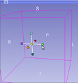
Incomplete Sphere, starting to scale
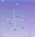
Incomplete Sphere, after scaling


Key Investigators
- University of Iowa: Christian Bauer, Reinhard Beichel, Markus Van Tol, ...?
Objective
We are developing a loadable module for Slicer to allow for threshold-based segmentation of tumors (or other regions of uptake) in PET scans within user-specified bounds and the tracking of such tumors. This would be used so that multiple physicians could segment the tumor and all consistently create one segmentation. With this, further work would go into checking values such as size and intensity values of tumors, which could be compared over time.
This project may become a part of another: http://www.na-mic.org/Wiki/index.php/2012_Summer_Project_Week:LongitudinalPETCTModule
Approach, Plan
Tasks, in order of importance/doability at this project week:
1. Set up module testing so it can expand as the rest of the module expands.
2. Start on the section for comparing segmentations in different images over time. (Would need information on relating data sets over time)
3. Debug issues with saving the scene and interacting with the Annotations module.
Progress
Before project week: Created a loadable module that currently allows threshold-based segmentation in user-defined regions of interest. Currently designed for PET scan images. Has functionality to suggest a threshold in a space and exclude other regions by uptake values or by user-defined excluded regions and points. Fully functional without the need to change to another module.
After project week:
Didn't end up working on things in approach. Instead, worked on creating a spherical region of interest (SROI) for annotations. This is currently partially done and reasonably functional, if not as apparent as desired.
Delivery Mechanism
This work will be delivered to the NA-MIC Kit as a (please select the appropriate options by noting YES against them below)
- ITK Module
- Slicer Module
- Built-in
- Extension -- commandline
- Extension -- loadable YES
- Other (Please specify)
References
- Reinhard Beichel's UIowa staff page: http://www.engineering.uiowa.edu/~rbeichel/index.html
- Quantitative Imaging Network (QIN) page: http://imaging.cancer.gov/programsandresources/specializedinitiatives/qin