Difference between revisions of "2014 Summer Project Week:Image To Mesh Conversion for Brain MRI"
m (Text replacement - "http://www.slicer.org/slicerWiki/index.php/" to "https://www.slicer.org/wiki/") |
|||
| (23 intermediate revisions by one other user not shown) | |||
| Line 1: | Line 1: | ||
| + | __NOTOC__ | ||
| + | <gallery> | ||
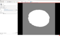
| + | Image:BCC_Slicer.png | BCC mesh of a brain labeled image | ||
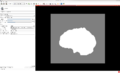
| + | Image:MC_Slicer.png | BCC mesh of a brain labeled image after MC | ||
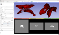
| + | Image:MeshBCCSection.jpg | BCC mesh (cross section) | ||
| + | Image:MeshBCCDeformedSection.jpg | BCC mesh after MC (cross section) | ||
| + | Image:BCCSlicerModule.jpg | BCC CLI module | ||
| + | Image:MCSlicerModule.jpg | MC CLI module | ||
| + | Image:NonConnectivity.jpg | How the Non-connectivity criterion influences the number of target points (MC module) | ||
| + | Image:Ventricles.jpg | A mesh of the brain ventricles after the MC. | ||
| + | </gallery> | ||
| + | |||
| + | ==Key Investigators== | ||
| + | * Fotis Drakopoulos (CRTC) | ||
| + | * Yixun Liu (CRTC) | ||
| + | * Andrey Fedorov (BWH/SPL) | ||
| + | * Ron Kikinis (BWH/SPL) | ||
| + | * Nikos Chrisochoides (CRTC) | ||
| + | |||
==Project Description== | ==Project Description== | ||
| − | + | The '''CBC3DI2MConversion''' project generates a tetrahedral mesh from an input labeled image. The method consists of two modules: | |
* Body Centric Cubic (BCC) Mesh Generation | * Body Centric Cubic (BCC) Mesh Generation | ||
| − | This | + | This module generates a Body Centric Cubic (BCC) mesh from a labeled image. Initially the generated mesh is homogeneous, that means does not distinguish different tissues. |
| − | Later the | + | Later the module specifies which tissue each tetrahedron belongs to. Each tissue is capable of automatically adjusting its resolution based on its geometric complexity and the |
predefined subdivision criterion. | predefined subdivision criterion. | ||
* Mesh Compression (MC) | * Mesh Compression (MC) | ||
| − | This | + | This module deforms a tetrahedral mesh towards the boundaries of the labeled image. Two point sets are extracted for the mesh deformation. The first (source point set) consists of the exterior surface vertices of the input mesh. The second (target point set) consists of the exterior surface edge points in the input labeled image. Then the input mesh is deformed by registering the source to the target point set using a Physics-Based Non-Rigid Registration method. |
<div style="margin: 20px;"> | <div style="margin: 20px;"> | ||
<div style="width: 27%; float: left; padding-right: 3%;"> | <div style="width: 27%; float: left; padding-right: 3%;"> | ||
<h3>Objective</h3> | <h3>Objective</h3> | ||
| − | * Develop | + | * Develop an extension that implements the Image-To-Mesh Conversion. |
| − | * | + | * The extension encapsulates two CLI modules: |
| + | ** BodyCentricCubic (BCC) mesh | ||
| + | ** Mesh Compression (MC) | ||
</div> | </div> | ||
<div style="width: 27%; float: left; padding-right: 3%;"> | <div style="width: 27%; float: left; padding-right: 3%;"> | ||
<h3>Approach, Plan</h3> | <h3>Approach, Plan</h3> | ||
| − | * | + | * The current version of the extension supports a single-tissue mesh generation. |
| + | * The future versions will support multi-tissue meshes. | ||
</div> | </div> | ||
<div style="width: 27%; float: left; padding-right: 3%;"> | <div style="width: 27%; float: left; padding-right: 3%;"> | ||
<h3>Progress</h3> | <h3>Progress</h3> | ||
| − | *The | + | * The experimental-build of the extension has uploaded on MIDAS dashboard (http://slicer.kitware.com/midas3/item/142306). |
| + | * The documentation page for the extension has created in the wiki (https://www.slicer.org/wiki/Documentation/Nightly/Extensions/ImageToMeshConversion). | ||
| + | * Three cases (brain, nidus and ventricles) are provided for testing. | ||
</div> | </div> | ||
</div> | </div> | ||
==References== | ==References== | ||
| − | *Tetrahedral | + | * Tetrahedral Mesh Generation for Medical Imaging. |
| − | Fedorov A., Chrisochoides N., Kikinis R., Warfield S. | + | Fedorov A., Chrisochoides N., Kikinis R., Warfield S., The Insight Journal - 2005 MICCAI Open-Source Workshop |
| − | *Mesh Deformation-based Multi-tissue Mesh Generation for Brain Images. | + | * Mesh Deformation-based Multi-tissue Mesh Generation for Brain Images. |
Yixun Liu, Panagiotis Foteinos, Andrey Chernikov and Nikos Chrisochoides. Engineering with Computers, Volume 28, pages 305-318, 2012. | Yixun Liu, Panagiotis Foteinos, Andrey Chernikov and Nikos Chrisochoides. Engineering with Computers, Volume 28, pages 305-318, 2012. | ||
| − | |||
| − | |||
| − | |||
| − | |||
| − | |||
| − | |||
| − | |||
Latest revision as of 18:07, 10 July 2017
Home < 2014 Summer Project Week:Image To Mesh Conversion for Brain MRI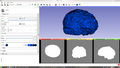
BCC mesh of a brain labeled image
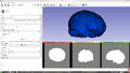
BCC mesh of a brain labeled image after MC
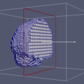
BCC mesh (cross section)
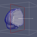
BCC mesh after MC (cross section)
BCC CLI module
MC CLI module

How the Non-connectivity criterion influences the number of target points (MC module)
A mesh of the brain ventricles after the MC.


Key Investigators
- Fotis Drakopoulos (CRTC)
- Yixun Liu (CRTC)
- Andrey Fedorov (BWH/SPL)
- Ron Kikinis (BWH/SPL)
- Nikos Chrisochoides (CRTC)
Project Description
The CBC3DI2MConversion project generates a tetrahedral mesh from an input labeled image. The method consists of two modules:
- Body Centric Cubic (BCC) Mesh Generation
This module generates a Body Centric Cubic (BCC) mesh from a labeled image. Initially the generated mesh is homogeneous, that means does not distinguish different tissues. Later the module specifies which tissue each tetrahedron belongs to. Each tissue is capable of automatically adjusting its resolution based on its geometric complexity and the predefined subdivision criterion.
- Mesh Compression (MC)
This module deforms a tetrahedral mesh towards the boundaries of the labeled image. Two point sets are extracted for the mesh deformation. The first (source point set) consists of the exterior surface vertices of the input mesh. The second (target point set) consists of the exterior surface edge points in the input labeled image. Then the input mesh is deformed by registering the source to the target point set using a Physics-Based Non-Rigid Registration method.
Objective
- Develop an extension that implements the Image-To-Mesh Conversion.
- The extension encapsulates two CLI modules:
- BodyCentricCubic (BCC) mesh
- Mesh Compression (MC)
Approach, Plan
- The current version of the extension supports a single-tissue mesh generation.
- The future versions will support multi-tissue meshes.
Progress
- The experimental-build of the extension has uploaded on MIDAS dashboard (http://slicer.kitware.com/midas3/item/142306).
- The documentation page for the extension has created in the wiki (https://www.slicer.org/wiki/Documentation/Nightly/Extensions/ImageToMeshConversion).
- Three cases (brain, nidus and ventricles) are provided for testing.
References
- Tetrahedral Mesh Generation for Medical Imaging.
Fedorov A., Chrisochoides N., Kikinis R., Warfield S., The Insight Journal - 2005 MICCAI Open-Source Workshop
- Mesh Deformation-based Multi-tissue Mesh Generation for Brain Images.
Yixun Liu, Panagiotis Foteinos, Andrey Chernikov and Nikos Chrisochoides. Engineering with Computers, Volume 28, pages 305-318, 2012.