Difference between revisions of "MRI-Guided Robot-assisted Deep Brain Stimulation Electrode PLacement"
From NAMIC Wiki
(Created page with 'Direct MR image guidance during deep brain stimulation (DBS) insertion offers many benefits; most significantly, interventional MRI can be used for planning, monitoring of tissue...') |
|||
| (12 intermediate revisions by the same user not shown) | |||
| Line 1: | Line 1: | ||
| − | + | __NOTOC__ | |
| + | <gallery> | ||
| + | Image:PW2009-v3.png|[[2009_Summer_Project_Week#Projects|Project Week Main Page]] | ||
| + | Image:MRNeuroRobot.jpg|Prototype Robot | ||
| + | Image:OpenIGTLink_Diagram_JHURobot.png|System Architecture based on CISST, Slicer and OpenIGTLInk | ||
| + | </gallery> | ||
| − | + | ==Key Investigators== | |
| − | + | * WPI: Gregory Fischer [mailto:gfischer@wpi.edu gfischer@wpi.edu] | |
| + | * UMass Med.: Julie Pilitsis, Mitch Albert | ||
| + | * Kitware: Michel Audette | ||
| + | |||
| + | ==Collaborators== | ||
| + | * JHU: Peter Kazanzides, Anton Deguet | ||
| + | * BWH: Junichi Tokuda, Noby Hata | ||
| + | * Kitware: Stephen Aylward, Andinet Enquobahrie | ||
| + | |||
| + | ==Students== | ||
| + | * WPI: Greg Cole (Robot, Motor Control), Hao Su (Haptics, Sensors), Yi Wang (Controller, Pneumatics), Kevin Harrington (Controller Software and Interface) | ||
| − | + | === Project Overview=== | |
| + | The objective of our research is to make conventional diagnostic closed high-field MRI scanners available for guiding deep brain stimulation electrode placement interventions. Our approach is to employ an MRI-compatible robotic assistant for guiding DBS electrode insertion under direct, real-time MR image guidance. The system will allow interactive probe alignment under real-time imaging in standard diagnostic high-field MR scanners. Use of a robotic assistant will minimize the potential for human error and mis-registration associated with the current procedure and will better address the practical issues of operating in an MR scanner bore. | ||
| − | == | + | === Project Week Plan === |
| − | * | + | |
| − | * | + | <div style="margin: 20px;"> |
| − | * | + | |
| + | <div style="width: 27%; float: left; padding-right: 3%;"> | ||
| + | <h3>Objective</h3> | ||
| + | * Robot control from Slicer | ||
| + | * Neuro navigation | ||
| + | * Electrode tracking in MRI | ||
| + | * Localization and tracking of brain anatomy (STN) | ||
| + | </div> | ||
| + | |||
| + | <div style="width: 27%; float: left; padding-right: 3%;"> | ||
| + | <h3>Approach, Plan</h3> | ||
| + | * Open to new collaborations to solve the listed objectives | ||
| + | </div> | ||
| − | = | + | <div style="width: 40%; float: left;"> |
| − | * | + | <h3>Progress</h3> |
| + | * Prototype robot assembled | ||
| + | * Modular MR robot controller ready | ||
| + | * Robot controller architecture using CISST functional | ||
| + | * Communication w/ OpenIGTLink functional | ||
| + | </div> | ||
| + | </div> | ||
| − | + | ==References== | |
| − | + | *Wang Y, Cole GA, Su H, Pilitis JG, Fischer GS, MRI Compatibility Evaluation of a Piezoelectric Actuator System for a Neural Interventional Robot, 31st Annual International Conference of the IEEE Engineering in Medicine and Biology Society - EMBC 2009, Minneapolis, Minnesota, September 2009 (accepted). | |
| − | + | *Cole G, Pilitsis J, Fischer GS, Design of a Robotic System for MRI-Guided Deep Brain Stimulation Electrode Placement, International Conference on Robotics and Automation - ICRA 2009, Kobe, Japan, May 2009. | |
| + | *Wang Y, Shazeeb MS, Sotak CH, Fischer GS, Optimization of Piezoelectric Motors to Enhance MR Compatiblity for Interventional Devices, 17th Scientific Meeting and Exhibition of the International Society of Magnetic Resonance in Medicine - ISMRM 2009, April 2009. | ||
Latest revision as of 19:10, 22 June 2009
Home < MRI-Guided Robot-assisted Deep Brain Stimulation Electrode PLacement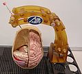
Prototype Robot
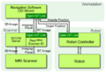
System Architecture based on CISST, Slicer and OpenIGTLInk


Key Investigators
- WPI: Gregory Fischer gfischer@wpi.edu
- UMass Med.: Julie Pilitsis, Mitch Albert
- Kitware: Michel Audette
Collaborators
- JHU: Peter Kazanzides, Anton Deguet
- BWH: Junichi Tokuda, Noby Hata
- Kitware: Stephen Aylward, Andinet Enquobahrie
Students
- WPI: Greg Cole (Robot, Motor Control), Hao Su (Haptics, Sensors), Yi Wang (Controller, Pneumatics), Kevin Harrington (Controller Software and Interface)
Project Overview
The objective of our research is to make conventional diagnostic closed high-field MRI scanners available for guiding deep brain stimulation electrode placement interventions. Our approach is to employ an MRI-compatible robotic assistant for guiding DBS electrode insertion under direct, real-time MR image guidance. The system will allow interactive probe alignment under real-time imaging in standard diagnostic high-field MR scanners. Use of a robotic assistant will minimize the potential for human error and mis-registration associated with the current procedure and will better address the practical issues of operating in an MR scanner bore.
Project Week Plan
Objective
- Robot control from Slicer
- Neuro navigation
- Electrode tracking in MRI
- Localization and tracking of brain anatomy (STN)
Approach, Plan
- Open to new collaborations to solve the listed objectives
Progress
- Prototype robot assembled
- Modular MR robot controller ready
- Robot controller architecture using CISST functional
- Communication w/ OpenIGTLink functional
References
- Wang Y, Cole GA, Su H, Pilitis JG, Fischer GS, MRI Compatibility Evaluation of a Piezoelectric Actuator System for a Neural Interventional Robot, 31st Annual International Conference of the IEEE Engineering in Medicine and Biology Society - EMBC 2009, Minneapolis, Minnesota, September 2009 (accepted).
- Cole G, Pilitsis J, Fischer GS, Design of a Robotic System for MRI-Guided Deep Brain Stimulation Electrode Placement, International Conference on Robotics and Automation - ICRA 2009, Kobe, Japan, May 2009.
- Wang Y, Shazeeb MS, Sotak CH, Fischer GS, Optimization of Piezoelectric Motors to Enhance MR Compatiblity for Interventional Devices, 17th Scientific Meeting and Exhibition of the International Society of Magnetic Resonance in Medicine - ISMRM 2009, April 2009.