Difference between revisions of "PA/A Tool"
From NAMIC Wiki
| (7 intermediate revisions by the same user not shown) | |||
| Line 2: | Line 2: | ||
<gallery> | <gallery> | ||
Image:PW-2015SLC.png|[[2015_Winter_Project_Week#Projects|Projects List]] | Image:PW-2015SLC.png|[[2015_Winter_Project_Week#Projects|Projects List]] | ||
| + | Image:Screen Shot 2015-01-05 at 15.09.46.png | Pulmonary Artery over Aorta ratio | ||
</gallery> | </gallery> | ||
| Line 11: | Line 12: | ||
<div style="width: 27%; float: left; padding-right: 3%;"> | <div style="width: 27%; float: left; padding-right: 3%;"> | ||
<h3>Objective</h3> | <h3>Objective</h3> | ||
| − | *Calculate in a fast way the ratio of the major and minor axis of the aorta in a chest CT slice where the pulmonary artery is clearly visible. This ratio is a biomarker used | + | *Calculate the ratio between the diameters of aorta and pulmonary artery, as a biomarker of acute exacerbations of COPD. |
| − | For more information about the medical need: | + | *Calculate in a fast way the ratio of the major and minor axis of the aorta in a chest CT slice where the pulmonary artery is clearly visible. This ratio is a biomarker used in pulmonary vascular disease and has been associated with outcomes like exacerbations. |
| − | + | For more information about the medical need: [http://www.ncbi.nlm.nih.gov/pmc/articles/PMC3690810/?tool=pmcentrez Pulmonary Arterial Enlargement and Acute Exacerbations of COPD] | |
</div> | </div> | ||
<div style="width: 27%; float: left; padding-right: 3%;"> | <div style="width: 27%; float: left; padding-right: 3%;"> | ||
<h3>Approach, Plan</h3> | <h3>Approach, Plan</h3> | ||
*Phase 1: | *Phase 1: | ||
| − | ** Give the expert a simple mecanism to measure the aorta | + | ** Give the expert a simple mecanism to measure the aorta and pulmonary artery is in the best slide possible (this could include an automatic tool to detect which is the best slide) |
| − | ** | + | ** Calculate the ratio automatically. |
* Phase 2: | * Phase 2: | ||
| − | ** Completely automatic | + | ** Completely automatic measurement of aorta and pumonary artery for an expert validation. |
</div> | </div> | ||
<div style="width: 27%; float: left; padding-right: 3%;"> | <div style="width: 27%; float: left; padding-right: 3%;"> | ||
<h3>Progress</h3> | <h3>Progress</h3> | ||
| − | * | + | * Markups are automatically set according to the structure that the user has selected |
| + | [[File:Aorta.png|300px]] | ||
</div> | </div> | ||
</div> | </div> | ||
Latest revision as of 01:49, 9 January 2015
Home < PA < A Tool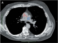
Pulmonary Artery over Aorta ratio


Key Investigators
Jorge Onieva, Rola Harmouche, German Gonzalez
Project Description
Objective
- Calculate the ratio between the diameters of aorta and pulmonary artery, as a biomarker of acute exacerbations of COPD.
- Calculate in a fast way the ratio of the major and minor axis of the aorta in a chest CT slice where the pulmonary artery is clearly visible. This ratio is a biomarker used in pulmonary vascular disease and has been associated with outcomes like exacerbations.
For more information about the medical need: Pulmonary Arterial Enlargement and Acute Exacerbations of COPD
Approach, Plan
- Phase 1:
- Give the expert a simple mecanism to measure the aorta and pulmonary artery is in the best slide possible (this could include an automatic tool to detect which is the best slide)
- Calculate the ratio automatically.
- Phase 2:
- Completely automatic measurement of aorta and pumonary artery for an expert validation.
Progress
- Markups are automatically set according to the structure that the user has selected
