Project Week 25/Breast Segmentation in DCE-MRI via Deep Learning Approaches
Back to Projects List
Breast Cancer Analysis in DCE-MRI
Key Investigators
- Gabriele Piantadosi (University Federico II di Napoli, Italy)\
Background
In recent years Dynamic Contrast Enhanced-Magnetic Resonance Imaging (DCE-MRI) has gained popularity as an important complementary diagnostic methodology for early detection of breast cancer. It has demonstrated a great potential in the screening of high-risk women, in staging newly diagnosed breast cancer patients and in assessing therapy effects thanks to its minimal invasiveness and to the possibility to visualise 3D high resolution dynamic (functional) information not available with conventional RX imaging. Among the major issues in developing CAD systems for breast DCE-MRI there are: (a) the detection of the suspicious region of interests (ROIs) as sensibly as possible, while simultaneously minimising the number of false alarms and (b) the classification of each segmented ROI according to its aggressiveness. This task is made harder by the peculiarity of DCE-MRI breast examinations: breast movements due to inspiration, a huge diversity of lesion types.
Project Description
Segmentation of the breast parenchyma could be approached as a classification problem.
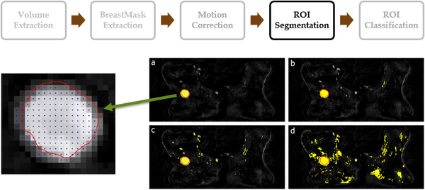
In the image, my previous proposal[1]: classical machine learning approaches using Support Vector Machine (SVM) trained with dynamic features, extracted from a suitable pre-selected area by using a pixel-based approach. A pre-selection mask strongly improved the final result.
The novel proposed lesion detection module performs the segmentation of lesions in Regions of Interest (ROIs) by means of classification at a pixel level or as dense regions relying on deep approaches such as those proposed in[2].
| Objective | Approach and Plan | Progress and Next Steps |
|---|---|---|
|
|
Relying on the segmented regions of interest (ROIs) a lesion malignity assessment via deep approaches should be performed. |
Background and References
- ↑ Marrone, S., Piantadosi, G., Fusco, R., Petrillo, A., Sansone, M., & Sansone, C. (2013, September). Automatic lesion detection in breast DCE-MRI. In International Conference on Image Analysis and Processing (pp. 359-368). Springer Berlin Heidelberg
- ↑ Long, J., Shelhamer, E., & Darrell, T. (2015). Fully convolutional networks for semantic segmentation. In Proceedings of the IEEE Conference on Computer Vision and Pattern Recognition (pp. 3431-3440)

