Difference between revisions of "SPIE 2012 DTI Workshop"
(→Venue) |
|||
| Line 4: | Line 4: | ||
[[Image:Spie2012 DTI course spujol.png|500px|right]] | [[Image:Spie2012 DTI course spujol.png|500px|right]] | ||
| − | The development of Diffusion Tensor Magnetic Resonance Imaging (DT-MRI) has opened up the possibility of studying the complex organization of the | + | The development of Diffusion Tensor Magnetic Resonance Imaging (DT-MRI) has opened up the possibility of studying the complex organization of the brain's white matter in-vivo. By measuring the diffusion of water molecules in tissues, the technique gives insights into the structure and orientation of major white matter pathways, and DT-MRI findings have the potential to play a critical role in the extraction of meaningful information for diagnosis, prognosis and following of treatment response. |
| − | The course will guide participants through the | + | |
| − | The hands-on sessions will use the | + | The course will guide participants through the fundamental aspects of DT-MRI data analysis, as well as the challenges of transferring cutting-edge DT-MRI techniques to clinical routine. The format will include a series of hands-on sessions with the participants running DT-MRI analysis on their own laptops, to provide a practical experience of extracting useful clinical information from Diffusion MR images. Participants will be guided through an integrated workflow for exploring the brain white matter in a series of datasets that will be provided as part of the course. The hands-on sessions will use DT-MRI tools from the NA-MIC toolkit, which include the 3DSlicer software, an open-source platform for medical image processing and 3D visualization used in biomedical and clinical research. |
| + | |||
| + | This event is part of the on-going effort of the NIH-funded National Alliance for Medical Image Computing (NA-MIC) to transfer the latest advances in biomedical image analysis to the scientific and clinical community. | ||
| + | |||
== Venue== | == Venue== | ||
| − | This is a workshop proposal for a Diffusion Tensor Imaging Workshop at [http://spie.org/x12166.xml SPIE Medical Imaging 2012, Feb.4-9, San Diego, California]. | + | This is a workshop proposal for a Hands On Diffusion Tensor Imaging Workshop at [http://spie.org/x12166.xml SPIE Medical Imaging 2012, Feb.4-9, San Diego, California]. |
==Faculty == | ==Faculty == | ||
| Line 26: | Line 29: | ||
== Learning Outcomes == | == Learning Outcomes == | ||
| − | + | This course will enable you to: | |
| − | * identify the different components of a | + | * identify the different components of a DT-MRI fiber tract analysis pipeline |
| − | * | + | * perform DWI/DTI data quality control |
| − | * generate white matter tracts in a normal subject and pathological case | + | * visualize 3D tensor fields and diffusion-derived maps |
| + | * generate 3D reconstructions of white matter tracts in a normal subject and pathological case | ||
| + | * extract and visualize DTI fiber tract profiles | ||
* identify the current challenges inherent in using DT-MRI data in the clinics | * identify the current challenges inherent in using DT-MRI data in the clinics | ||
| + | |||
== Intended Audience == | == Intended Audience == | ||
Scientists, engineers, and clinical researchers who are interested in learning how to use Diffusion Tensor MR Imaging for mapping the white matter of the human brain in health and disease. | Scientists, engineers, and clinical researchers who are interested in learning how to use Diffusion Tensor MR Imaging for mapping the white matter of the human brain in health and disease. | ||
Revision as of 13:07, 14 October 2011
Home < SPIE 2012 DTI Workshop(Note: This a proposal under consideration.)
Contents
Course Description
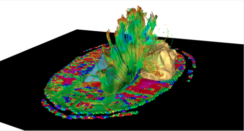
The development of Diffusion Tensor Magnetic Resonance Imaging (DT-MRI) has opened up the possibility of studying the complex organization of the brain's white matter in-vivo. By measuring the diffusion of water molecules in tissues, the technique gives insights into the structure and orientation of major white matter pathways, and DT-MRI findings have the potential to play a critical role in the extraction of meaningful information for diagnosis, prognosis and following of treatment response.
The course will guide participants through the fundamental aspects of DT-MRI data analysis, as well as the challenges of transferring cutting-edge DT-MRI techniques to clinical routine. The format will include a series of hands-on sessions with the participants running DT-MRI analysis on their own laptops, to provide a practical experience of extracting useful clinical information from Diffusion MR images. Participants will be guided through an integrated workflow for exploring the brain white matter in a series of datasets that will be provided as part of the course. The hands-on sessions will use DT-MRI tools from the NA-MIC toolkit, which include the 3DSlicer software, an open-source platform for medical image processing and 3D visualization used in biomedical and clinical research.
This event is part of the on-going effort of the NIH-funded National Alliance for Medical Image Computing (NA-MIC) to transfer the latest advances in biomedical image analysis to the scientific and clinical community.
Venue
This is a workshop proposal for a Hands On Diffusion Tensor Imaging Workshop at SPIE Medical Imaging 2012, Feb.4-9, San Diego, California.
Faculty
- Sonia Pujol, Ph.D., Surgical Planning Laboratory, Brigham and Women’s Hospital, Harvard Medical School
- Martin Styner, Ph.D.,Neuro Image Research and Analysis Laboratory, University of North Carolina
- Guido Gerig, Ph.D., The Scientific Computing and Imaging Institute, University of Utah
Tentative Agenda
- 5:45-5:50 pm: Introduction and goals of the workshop (Sonia Pujol)
- 5:50-6:25 pm: Diffusion tensor processing and visualization: getting to know DTI data very, very well- tensors, glyphs and more (Guido Gerig)
- 6:25-7:00 pm: DTI in research and in the clinics: current uses and future roadmap (Martin Styner)
- 7:00-7:30 pm: Challenges in clinical transfer of DT-MRI: towards validation of tractography (Sonia Pujol)
- 7:30-7:50 pm: Hands-on session: White matter exploration for neurosurgical planning (Sonia Pujol)
- 7:50-8:00 pm: Concluding remarks and discussion
Learning Outcomes
This course will enable you to:
- identify the different components of a DT-MRI fiber tract analysis pipeline
- perform DWI/DTI data quality control
- visualize 3D tensor fields and diffusion-derived maps
- generate 3D reconstructions of white matter tracts in a normal subject and pathological case
- extract and visualize DTI fiber tract profiles
- identify the current challenges inherent in using DT-MRI data in the clinics
Intended Audience
Scientists, engineers, and clinical researchers who are interested in learning how to use Diffusion Tensor MR Imaging for mapping the white matter of the human brain in health and disease.
