ProjectWeek200706:LesionClassificationInLupus
From NAMIC Wiki
(Redirected from Projects/Structural/2007 Project Week Lesion Classification in Lupus)
Home < Projects < Structural < 2007 Project Week Lesion Classification in Lupus
 Return to Project Week Main Page |
Key Investigators
- MIND/UNM: Jeremy Bockholt, Mark Scully
- MGH:Bruce Fischl
- UIowa: Vincent Magnotta
Objective
Our goal is to automatically, or with little or no manual human rater input, accurately tissue classify our example lupus data-set into gray, white, csf, and lesion classes.
Approach, Plan
Our approach is to utilize existing automatic lesion classification techniques, both within NA-MIC kit and external to it.
Our plan for the project week is to learn and use EMSegment (S. Wells), BRAINS2 (V. Magnotta), and Constructing Image Graphs for Lesion Segmention (M. Prastawa). We will learn and begin to compare and contrast these approaches with a bronze standard (manual rater tracings) of lesions.
Progress
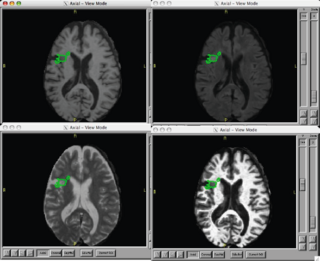
- Provided Kilian Pohl and Brad Davis an example anonymous demo case with raw t1, t2, and flair, and coregistered t1, t2, flair and manually traced lesion images.
- Mark Scully has installed current Slicer3 and run the EMSegment tutorial with tutorial data, we sucessfully ran the stock EMSegment Slicer3 tutorial, and then modified the demo case in order to see how we would run our lesion data
- Kilian and Brad helped us with several Slicer3 EMSegment questions
- We talked with Brad about the need of the EMSegment module (or other module in Slicer) to provide co-registration of t1, t2, and flair, as well as registration of input modalities to the atlas images
- At the moment we are still working on getting our images registered to the atlas using FSL
- We talked with Vince Magnotta about how to run the BRAINS2 lesion method on this example lupus data
- Mark came up to speed on ITK/VTK and will be comfortable working with the NA-MIC kit
- Future Worklist
- Share data with Guido to apply the Image Graphs for Lesion Segmention (M. Prastawa) approach on this sample data set
References
Additional Information
Using an exemplar case that has already been processed using non-NAMIC kit tools:
- Use existing NA-MIC kit to coregister T1, T2, Flair
- Use the EM Segment in slicer 2-3.X to classify grey, white, csf, and white matter lesion
- Summarize the volume and location of lesions
