Difference between revisions of "Leadership:RKBiomedComp2009"
From NAMIC Wiki
| (12 intermediate revisions by the same user not shown) | |||
| Line 1: | Line 1: | ||
| + | __NOTOC__ | ||
| + | [[Leadership:Main|Back to Leadership:Main]] | ||
=Lecture on 10-01-2009= | =Lecture on 10-01-2009= | ||
| Line 4: | Line 6: | ||
=Image acquisition= | =Image acquisition= | ||
| + | |||
| + | {| | ||
| + | |align="left"|This is a listing of some of the medical imaging modalities which are used routinely in clinical practice in the the US. | ||
*[http://en.wikipedia.org/wiki/X-ray X-rays] | *[http://en.wikipedia.org/wiki/X-ray X-rays] | ||
*[http://en.wikipedia.org/wiki/Ultrasound Ultrasound] | *[http://en.wikipedia.org/wiki/Ultrasound Ultrasound] | ||
| Line 9: | Line 14: | ||
*[http://en.wikipedia.org/wiki/Nuclear_medicine Nuclear Medicine: Spect, Pet] | *[http://en.wikipedia.org/wiki/Nuclear_medicine Nuclear Medicine: Spect, Pet] | ||
*[http://en.wikipedia.org/wiki/MRI MRI: Structural, functional, DTI] | *[http://en.wikipedia.org/wiki/MRI MRI: Structural, functional, DTI] | ||
| + | *[http://en.wikipedia.org/wiki/Medical_imaging#Creation_of_three-dimensional_images Postprocessing] | ||
| + | | | ||
| + | {| | ||
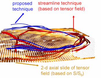
| + | |+ '''Example of MR tractography analysis''' | ||
| + | |valign="top"|[[Image:Tracts2.png|thumb|350px|See [[Algorithm:GATech:Finsler_Active_Contour_DWI|here]] for more information]] | ||
| + | |} | ||
| + | |} | ||
=Image Informatics= | =Image Informatics= | ||
| Line 14: | Line 26: | ||
=Medical Image Computing= | =Medical Image Computing= | ||
| − | [[media:Kikinis-IIC-10-03-2007.ppt|IIC 2007 presentation]] | + | {| |
| − | [[media:2009-06-18-Kikinis-CAOS.ppt|CAOS 2009 presentation]] | + | |align="left"| The following two powerpoint presentations provide an introduction to medical image computing, as practiced at the SPL, and to some basic concepts used for image guided therapy (IGT). |
| + | *[[media:Kikinis-IIC-10-03-2007.ppt|IIC 2007 presentation]]: MIC | ||
| + | *[[media:2009-06-18-Kikinis-CAOS.ppt|CAOS 2009 presentation]]: IGT | ||
| + | *[http://www.slicer.org Slicer website] | ||
| + | | | ||
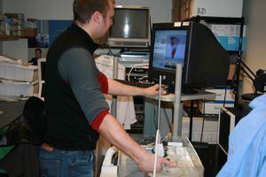
| + | [[Image:US_Photoseries_4.png|300px|thumb|4D ultrasound imaging within Slicer3. Paul Novotny, Children's Hospital Boston, Boston and Ben Grauer, Swiss Federal Inst of Tech.]] | ||
| + | |} | ||
Latest revision as of 11:58, 1 October 2009
Home < Leadership:RKBiomedComp2009Lecture on 10-01-2009
See here for a schedule of the entire 6.872J/HST950 Biomedical Computing class.
Image acquisition
| This is a listing of some of the medical imaging modalities which are used routinely in clinical practice in the the US. |
|
Image Informatics
PACS systems: XNAT
Medical Image Computing
| The following two powerpoint presentations provide an introduction to medical image computing, as practiced at the SPL, and to some basic concepts used for image guided therapy (IGT). |

