Difference between revisions of "Projects:RegistrationLibrary:RegLib C12"
From NAMIC Wiki
| Line 39: | Line 39: | ||
[ | [ | ||
===Download === | ===Download === | ||
| − | *'''[[Media:RegLib_C12_LiverTumor_DATA.zip|download input image data <small> (Input Data, NRRD images, zip file | + | *'''[[Media:RegLib_C12_LiverTumor_DATA.zip|download input image data <small> (Input Data, NRRD images, zip file 42 MB) </small>]]''' |
*'''[[Media:RegLib_C12_LiverTumorAblation1_ParameterPresets.mrml|download registration parameter presets file <small> (.mrml file 20 kB) </small>]]''' | *'''[[Media:RegLib_C12_LiverTumorAblation1_ParameterPresets.mrml|download registration parameter presets file <small> (.mrml file 20 kB) </small>]]''' | ||
*'''[[Media:RegLib_C12_LiverTumorAblation1_Tutorial.ppt|download guided tutorial <small> (PowerPoint, xx MB) </small>]]''' | *'''[[Media:RegLib_C12_LiverTumorAblation1_Tutorial.ppt|download guided tutorial <small> (PowerPoint, xx MB) </small>]]''' | ||
Revision as of 16:38, 2 March 2010
Home < Projects:RegistrationLibrary:RegLib C12Back to ARRA main page
Back to Registration main page
Back to Registration Use-case Inventory
Contents
Slicer Registration Use Case Exampe #12: Liver Tumor Cryoablation
Objective / Background
We seek to align the pre-operative CT with the intra-operative MRI.
Keywords
MRI, CT, IGT, intra-operative, liver, cryoablation, change detection, non-rigid registration
Input Data
 reference/fixed : intra-op MRI, 0.78 x 0.78 x 2.5 mm axial, RAS orientation.
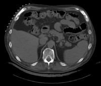
 moving: pr-op CT, 0.95 x 0.95 x 5 mm voxel size, axial, RAS orientation.
Methods
Registration Results
[
Download
- download input image data (Input Data, NRRD images, zip file 42 MB)
- download registration parameter presets file (.mrml file 20 kB)
- download guided tutorial (PowerPoint, xx MB)
- download full tutorial set (Input Data, presets, results, tutorial, zip file xx MB)
Link to User Guide: How to Load/Save Registration Parameter Presets
Discussion: Registration Challenges
- large differences in FOV
- strong differences in image contrast between MRI & CT
- contrast enhancement and pathology and treatment changes cause additional differences in image content
- we have strongly anisotropic voxel sizes with much less through-plane resolution


