Difference between revisions of "Projects:RegistrationLibrary:RegLib C19"
From NAMIC Wiki
| Line 60: | Line 60: | ||
*We have two separate sets of registrations to combine. While theoretically possible to simply align every scan with the reference directly, this is likely to produce inferior results. Basic rule of thumb is to register to the image that is closest in anatomy and contrast, in that order. | *We have two separate sets of registrations to combine. While theoretically possible to simply align every scan with the reference directly, this is likely to produce inferior results. Basic rule of thumb is to register to the image that is closest in anatomy and contrast, in that order. | ||
*the MRA has a strongly clipped FOV and low tissue contrast | *the MRA has a strongly clipped FOV and low tissue contrast | ||
| + | *Currently (Slicer 3.6) the concatenation of linear and nonlinear transforms is not (yet) supported directly in slicer. However this is an easy and fast procedure that can be done with any text editor. We merely copy the affine portion from one transform file and paste it into the nonrigid transform file. | ||
=== Discussion: Key Strategies === | === Discussion: Key Strategies === | ||
Revision as of 15:20, 2 July 2010
Home < Projects:RegistrationLibrary:RegLib C19Back to ARRA main page
Back to Registration main page
Back to Registration Use-case Inventory
Slicer Registration Library Exampe #19: Multi-contrast group analysis: intra- and inter-subject registration of multi-contrast MRI
Objective / Background
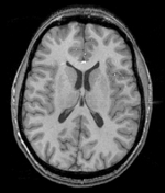
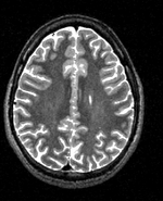
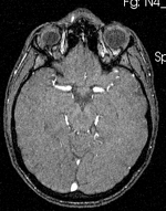
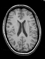
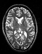
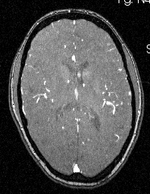
This is an example of multi-stage registration containing both intra- and inter-subject alignment. We have two sets of multi-sequence MRI for two subjects, each comprised of a T1, T2 and perfusion MRA scan. We ant everything aligned to a single space to enable regional or voxel-based group comparison.
Keywords
multi-stage registration, MRI, brain, head, multi-contrast, inter-subject, group analysis, MRA, T2
Methods
- align T2 and MRA of each subject to their respective T1 via an affine transform
- align the T1 of subject 2 to the T1 of subject 1 via a non-rigid transform, this establishes the inter-subject mapping
- combine the affine transforms of subject 2 T2 and MRA with the above nonrigid and resample the two images into the reference space
Registration Results
Download
- download image data (Original Data, Result transforms, parameter presets, zip file 76 MB)
- download guided tutorial (power point file, 1.5 MB)
- download registration parameter presets (mrml file, 12 kB)
Link to User Guide: How to Load/Save Registration Parameter Presets
Discussion: Registration Challenges
- We have two separate sets of registrations to combine. While theoretically possible to simply align every scan with the reference directly, this is likely to produce inferior results. Basic rule of thumb is to register to the image that is closest in anatomy and contrast, in that order.
- the MRA has a strongly clipped FOV and low tissue contrast
- Currently (Slicer 3.6) the concatenation of linear and nonlinear transforms is not (yet) supported directly in slicer. However this is an easy and fast procedure that can be done with any text editor. We merely copy the affine portion from one transform file and paste it into the nonrigid transform file.
Discussion: Key Strategies
- For the intra-subject affine portion: BRAINSfit
- For the inter-subject nonrigid portion: Fast nonrigid BSpline
- For combining the transforms: copy and paste in a Text Editor
- For applying the transforms and resampling: Resample Scalar/Vector/DWI Volume
Acknowledgments
- dataset provided by the
