Difference between revisions of "RSNA 3D Visualization Course"
(Created page with 'right|300 px The '''3D Visualization of DICOM Images for Radiological Applications''' course will be offered by the Neuroimage Analysis Center (…') |
|||
| Line 1: | Line 1: | ||
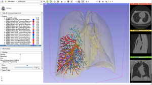
[[image:Fig5_ChestCT_Slicer.png|right|300 px]] | [[image:Fig5_ChestCT_Slicer.png|right|300 px]] | ||
| − | The '''3D Visualization of DICOM Images for Radiological Applications''' course will be offered by the Neuroimage Analysis Center (NAC), at the [http://rsna2014.rsna.org 100th Scientific Assembly and Annual Meeting of the Radiological Society of North America (RSNA 2014)]. As part of the outreach mission of this NIH funded National Center, we have developed an offering of freely available, multi-platforms open source software to enable medical image analysis research. The 3D Visualization course ([[media:3DVisualizationDICOM_SoniaPujol_RSNA2013.pdf |PDF]] or [[media:3DVisualizationDICOM_SoniaPujol_RSNA2013.pptx |PowerPoint]]) along with the [[Media:3DVisualization DICOM images part1.zip|3D Visualization-part1]] and [[Media:3DVisualization DICOM images part2.zip|3D Visualization-part2]] datasets aim to introduce translational clinical scientists to the basics of viewing and interacting in 3D with DICOM volumes and anatomical models using the 3DSlicer software. The version of the software that will be used during this course is Slicer4.4 and it can be downloaded [http://slicer.kitware.com/midas3/folder/274 here]. | + | The '''[http://rsna2014.rsna.org/pdf/11033051.pdf 3D Visualization of DICOM Images for Radiological Applications]''' course will be offered by the Neuroimage Analysis Center (NAC), at the [http://rsna2014.rsna.org 100th Scientific Assembly and Annual Meeting of the Radiological Society of North America (RSNA 2014)]. As part of the outreach mission of this NIH funded National Center, we have developed an offering of freely available, multi-platforms open source software to enable medical image analysis research. The 3D Visualization course ([[media:3DVisualizationDICOM_SoniaPujol_RSNA2013.pdf |PDF]] or [[media:3DVisualizationDICOM_SoniaPujol_RSNA2013.pptx |PowerPoint]]) along with the [[Media:3DVisualization DICOM images part1.zip|3D Visualization-part1]] and [[Media:3DVisualization DICOM images part2.zip|3D Visualization-part2]] datasets aim to introduce translational clinical scientists to the basics of viewing and interacting in 3D with DICOM volumes and anatomical models using the 3DSlicer software. The version of the software that will be used during this course is Slicer4.4 and it can be downloaded [http://slicer.kitware.com/midas3/folder/274 here]. |
For additional training materials on the software, please visit the [http://www.slicer.org/slicerWiki/index.php/Documentation/4.3/Training 3D Slicer Compendium]. | For additional training materials on the software, please visit the [http://www.slicer.org/slicerWiki/index.php/Documentation/4.3/Training 3D Slicer Compendium]. | ||
*CME Content Code: Informatics | *CME Content Code: Informatics | ||
Revision as of 19:00, 19 November 2014
Home < RSNA 3D Visualization CourseThe 3D Visualization of DICOM Images for Radiological Applications course will be offered by the Neuroimage Analysis Center (NAC), at the 100th Scientific Assembly and Annual Meeting of the Radiological Society of North America (RSNA 2014). As part of the outreach mission of this NIH funded National Center, we have developed an offering of freely available, multi-platforms open source software to enable medical image analysis research. The 3D Visualization course (PDF or PowerPoint) along with the 3D Visualization-part1 and 3D Visualization-part2 datasets aim to introduce translational clinical scientists to the basics of viewing and interacting in 3D with DICOM volumes and anatomical models using the 3DSlicer software. The version of the software that will be used during this course is Slicer4.4 and it can be downloaded here. For additional training materials on the software, please visit the 3D Slicer Compendium.
- CME Content Code: Informatics
- CME Credits Categories:
- AMA PRA Category 1 Credits™: 1.5
- ARRT Category A+ Credit: 1.5
Teaching Faculty
- Sonia Pujol, Ph.D., Surgical Planning Laboratory, Department of Radiology, Brigham and Women’s Hospital, Harvard Medical School, Boston MA.
- Ron Kikinis, M.D., Surgical Planning Laboratory, Department of Radiology, Brigham and Women’s Hospital, Harvard Medical School, Boston MA.
- Kitt Shaffer, M.D. Ph.D., Boston University School of Medicine, Department of Radiology, Boston Medical Center, Boston, MA.
Logistics
- Date: Monday December 1, 12:30-2:00 pm
- Location: Room S401AB, McCormick Conference Center, Chicago, IL.
- Registration: Sold-out
