Difference between revisions of "2016 Summer Project Week/Brain atlas combining histology and MRI"
(Edit "key investigators") |
|||
| Line 1: | Line 1: | ||
| + | <gallery> | ||
| + | Image:PW-Summer2016.png|[[2016_Summer_Project_Week#Projects|Projects List]] | ||
| + | Image:Brainstem contours on different images.png|Manually segmented structures of a human brainstem superimposed on a histological slice, a block-face volume and a 11.7 MRI. | ||
| + | Image:Brainstem contours 3D.png|3D visualization of the manually traced contours that have been used to interpolate the masks of the structures. | ||
| + | </gallery> | ||
| + | |||
==Key Investigators== | ==Key Investigators== | ||
* Fernando Pérez García ([http://icm-institute.org/en/ Institute of the Brain and Spine], France) | * Fernando Pérez García ([http://icm-institute.org/en/ Institute of the Brain and Spine], France) | ||
Revision as of 09:10, 24 June 2016
Home < 2016 Summer Project Week < Brain atlas combining histology and MRI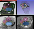
Manually segmented structures of a human brainstem superimposed on a histological slice, a block-face volume and a 11.7 MRI.
3D visualization of the manually traced contours that have been used to interpolate the masks of the structures.


Key Investigators
- Fernando Pérez García (Institute of the Brain and Spine, France)
- Sara Fernandez Vidal (Institute of the Brain and Spine, France)
- Sonia Pujol (BWH)
- Steve Pieper
- Csaba Pinter
- Andras Lasso
Project Description
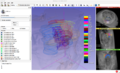
Multimodal 3D visualization is important during the construction of a 3D histology / MRI atlas. Also, the interface must be very intuitive so that the user can easily trace the many contours needed for a precise 3D reconstruction.
We have chosen 3D Slicer as our development platform because of its flexibility and its huge potential for multimodal 3D visualization. Our goal is to develop an integrated, intuitive extension for manual tracing of contours on stained histological slices that have been coregistered to a high resolution MRI.
| Objective | Approach and Plan | Progress and Next Steps |
|---|---|---|
|
|