Difference between revisions of "2009 Winter Project Week Gofigure LevelSet"
From NAMIC Wiki
Agouaillard (talk | contribs) (New page: {| |thumb|320px|Return to [[2009_Winter_Project_Week|Project Week Main Page ]] |[[]] |[[]] |} __NOTOC__ ===Key Investigators=== * kishore mosanliganti, Harvard ...) |
|||
| (6 intermediate revisions by the same user not shown) | |||
| Line 1: | Line 1: | ||
{| | {| | ||
|[[Image:NAMIC-SLC.jpg|thumb|320px|Return to [[2009_Winter_Project_Week|Project Week Main Page]] ]] | |[[Image:NAMIC-SLC.jpg|thumb|320px|Return to [[2009_Winter_Project_Week|Project Week Main Page]] ]] | ||
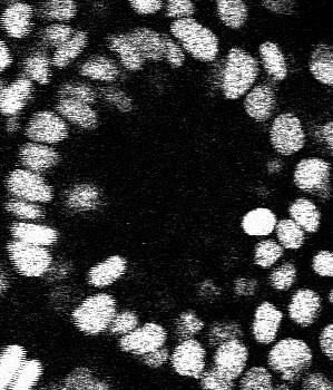
| − | |[[]] | + | |[[Image:GoFigureZebrafish2D.png|thumb|320px|Return to [[2009_Winter_Project_Week|Project Week Main Page]] ]] |
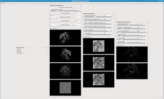
| − | |[[]] | + | |[[Image:KwWidgetLevelSetSegmentationGUI.png|thumb|320px|Return to [[2009_Winter_Project_Week|Project Week Main Page]] ]] |
|} | |} | ||
| Line 10: | Line 10: | ||
===Key Investigators=== | ===Key Investigators=== | ||
| − | * | + | * Kishore Mosaliganti, Harvard Medical School |
* Arnaud Gelas, Harvard Medical School | * Arnaud Gelas, Harvard Medical School | ||
* Alexandre Gouaillard, Harvard Medical School | * Alexandre Gouaillard, Harvard Medical School | ||
| + | * Luca Antiga, Mario Negri Institute | ||
| + | * Luis Ibanez, Kitware | ||
| Line 20: | Line 22: | ||
<h1>Objective</h1> | <h1>Objective</h1> | ||
| − | * Implement references 1, | + | * Implement a 3D+t image analysis pipeline for multistain microscopy images |
| − | * | + | * Use references 1-4 for preprocessing, cell segmentation and tracking in ITK |
| − | + | * Make a submission to Insight Journal | |
| Line 30: | Line 32: | ||
<h1>Approach, Plan</h1> | <h1>Approach, Plan</h1> | ||
| − | * | + | * First prototype of filters are already implemented |
| − | * Code | + | * Code review to be done with an ITK expert (luis/bill/jim?) |
| − | * Run on cell datasets | + | * Run on cell datasets and build relevant tests |
* Write IJ paper | * Write IJ paper | ||
| − | |||
</div> | </div> | ||
| Line 41: | Line 42: | ||
<h1>Progress</h1> | <h1>Progress</h1> | ||
| − | + | * Improved code quality by making it more memory and run-time efficient | |
| + | * Wrote test scripts and examples | ||
| + | * Run on cell datasets and exchanged code with Shantanu, OSU and Luca | ||
| + | * Began writing IJ paper | ||
</div> | </div> | ||
| Line 50: | Line 54: | ||
===References=== | ===References=== | ||
| + | # A. Hyvärinen. Fast and Robust Fixed-Point Algorithms for Independent Component Analysis, IEEE Transactions on Neural Networks 10(3):626-634, 1999. | ||
| + | # M. Leventon, W. Grimson and O. Faugeras. Statistical shape influence in geodesic active contours, Computer Vision and Pattern Recognition, 1, pp. 316-323, 2000. | ||
| + | # Tony Chan and Luminita Vese. An active contour model without edges, International Conference on Scale-Space Theories in Computer Vision, pp. 141-151, 1999. | ||
# Kishore Mosaliganti, Lee Cooper, Richard Sharp, Raghu Machiraju, Gustavo Leone, Kun Huang and Joel Saltz. Reconstruction of Cellular Biological Structures from Optical Microscopy Data. In IEEE Transactions in Visualization and Computer Graphics, 14 (4), pp. 863-876, July/August 2008. | # Kishore Mosaliganti, Lee Cooper, Richard Sharp, Raghu Machiraju, Gustavo Leone, Kun Huang and Joel Saltz. Reconstruction of Cellular Biological Structures from Optical Microscopy Data. In IEEE Transactions in Visualization and Computer Graphics, 14 (4), pp. 863-876, July/August 2008. | ||
# Tensor Classification of N-point Correlation Function features for Histology Tissue Segmentation. K. Mosaliganti, F. Janoos, O. Irfanoglu, R. Ridgway, R. Machiraju, K. Huang, J. Saltz, G. Leone and M. Ostrowski. Special Issue on Medical Image Analysis with Applications in Biology, Journal of Medical Image Analysis, 2008. | # Tensor Classification of N-point Correlation Function features for Histology Tissue Segmentation. K. Mosaliganti, F. Janoos, O. Irfanoglu, R. Ridgway, R. Machiraju, K. Huang, J. Saltz, G. Leone and M. Ostrowski. Special Issue on Medical Image Analysis with Applications in Biology, Journal of Medical Image Analysis, 2008. | ||
# Gang Li, Tianming Liu, Ashley Tarokh, Jingxin Nie, Lei Guo, Andrew Mara, Scott Holley and Stephen TC Wong. 3D cell nuclei segmentation based on gradient flow tracking. BMC Cell Biology, 8:40, 2007. | # Gang Li, Tianming Liu, Ashley Tarokh, Jingxin Nie, Lei Guo, Andrew Mara, Scott Holley and Stephen TC Wong. 3D cell nuclei segmentation based on gradient flow tracking. BMC Cell Biology, 8:40, 2007. | ||
Latest revision as of 16:33, 9 January 2009
Home < 2009 Winter Project Week Gofigure LevelSet Return to Project Week Main Page |
 Return to Project Week Main Page |
 Return to Project Week Main Page |
Key Investigators
- Kishore Mosaliganti, Harvard Medical School
- Arnaud Gelas, Harvard Medical School
- Alexandre Gouaillard, Harvard Medical School
- Luca Antiga, Mario Negri Institute
- Luis Ibanez, Kitware
Objective
- Implement a 3D+t image analysis pipeline for multistain microscopy images
- Use references 1-4 for preprocessing, cell segmentation and tracking in ITK
- Make a submission to Insight Journal
Approach, Plan
- First prototype of filters are already implemented
- Code review to be done with an ITK expert (luis/bill/jim?)
- Run on cell datasets and build relevant tests
- Write IJ paper
Progress
- Improved code quality by making it more memory and run-time efficient
- Wrote test scripts and examples
- Run on cell datasets and exchanged code with Shantanu, OSU and Luca
- Began writing IJ paper
References
- A. Hyvärinen. Fast and Robust Fixed-Point Algorithms for Independent Component Analysis, IEEE Transactions on Neural Networks 10(3):626-634, 1999.
- M. Leventon, W. Grimson and O. Faugeras. Statistical shape influence in geodesic active contours, Computer Vision and Pattern Recognition, 1, pp. 316-323, 2000.
- Tony Chan and Luminita Vese. An active contour model without edges, International Conference on Scale-Space Theories in Computer Vision, pp. 141-151, 1999.
- Kishore Mosaliganti, Lee Cooper, Richard Sharp, Raghu Machiraju, Gustavo Leone, Kun Huang and Joel Saltz. Reconstruction of Cellular Biological Structures from Optical Microscopy Data. In IEEE Transactions in Visualization and Computer Graphics, 14 (4), pp. 863-876, July/August 2008.
- Tensor Classification of N-point Correlation Function features for Histology Tissue Segmentation. K. Mosaliganti, F. Janoos, O. Irfanoglu, R. Ridgway, R. Machiraju, K. Huang, J. Saltz, G. Leone and M. Ostrowski. Special Issue on Medical Image Analysis with Applications in Biology, Journal of Medical Image Analysis, 2008.
- Gang Li, Tianming Liu, Ashley Tarokh, Jingxin Nie, Lei Guo, Andrew Mara, Scott Holley and Stephen TC Wong. 3D cell nuclei segmentation based on gradient flow tracking. BMC Cell Biology, 8:40, 2007.