Difference between revisions of "NA-MIC External Collaborations"
m (Text replacement - "http://www.slicer.org/slicerWiki/index.php/" to "https://www.slicer.org/wiki/") |
|||
| (40 intermediate revisions by 7 users not shown) | |||
| Line 1: | Line 1: | ||
| − | Back to [[NA-MIC_Collaborations|NA-MIC Collaborations]] | + | Back to [[NA-MIC_Collaborations|NA-MIC Collaborations]] |
| − | + | __TOC__ | |
=Projects funded by "Collaborations with NCBC PAR"= | =Projects funded by "Collaborations with NCBC PAR"= | ||
| − | This section describes external collaborations with NA-MIC that are funded by NIH under the "Collaboration with NCBC" PAR. (Details for this funding mechanism are provided [[Collaborator:Resources| | + | This section describes external collaborations with NA-MIC that are funded by NIH under the "Collaboration with NCBC" PAR. (Details for this funding mechanism are provided [[Collaborator:Resources|'''here''']]). |
| − | {| | + | {| class="wikitable" |
| − | | style=" | + | |- |
| − | | | + | |style="background: white"|[[Image:MichiganTMJ.png|216px]] |
| + | |[[PAR-12-001: R01DE024450 University of Michigan Quantification Of 3d Bony Changes In Temporomandibular Joint Osteoarthritis]]<br>The group at Michigan is working to precisely quantify three-dimensional bony changes in the Temporomandibular joint with the goal of improved early diagnosis and better monitoring of treatment outcomes. | ||
| + | [http://projectreporter.nih.gov/project_info_description.cfm?aid=8576556&icde=18353487 NIH Reporter Abstract] | ||
| − | = | + | Funding Duration: '''09/10/2013-08/31/2017''' |
| + | |- | ||
| + | |style="background: white"|[[Image:JHUSkullStripping.png|216px]]<br> [[Image:JHU.jpg]] | ||
| + | |[[NA-MIC_JHU_Skull_Stripping_Collaboration | PAR-08-183: R21EB009900 Johns Hopkins Skull Stripping]]<br>The group at Johns Hopkins is developing software that enables the stripping of skull, scalp, and meninges from structural MRI scans in a fully automated fashion. | ||
| − | + | [http://projectreporter.nih.gov/project_info_description.cfm?aid=7922035&icde=7494438 NIH Reporter Abstract] | |
| + | Funding Duration: '''08/01/2009-07/31/2011''' | ||
|- | |- | ||
| + | |[[Image:NhpATOCPic.jpg|216px]] | ||
| + | |[[NA-MIC_NCBC_Collaboration:Measuring_Alcohol_and_Stress_Interaction|PAR-07-249: R01AA016748 Measuring Alcohol and Stress Interaction with Structural and Perfusion MRI]]<br>This project is funded under an NCBC collaboration grant to PIs James Daunais, Robert Kraft, and Chris Wyatt. The goal of this project is to examine the the effects of chronic alcohol self-administration on brain structure and function the monkey brain. MRI image analysis tools from the [[NA-MIC-Kit|NA-MIC kit]] will be adapted for use with the monkey brain datasets. [[NA-MIC_NCBC_Collaboration:Measuring_Alcohol_and_Stress_Interaction|More...]] | ||
| − | + | [http://projectreporter.nih.gov/project_info_description.cfm?aid=7599715&icde=7511346 NIH Reporter Abstract] | |
| − | |||
| − | |||
| − | |||
| − | |||
| − | |||
| + | Funding Duration: '''07/15/2009-03/31/2010''' | ||
|- | |- | ||
| + | |[[Image:JHUCollaboration.jpg|216px]] | ||
| + | |[[NA-MIC_NCBC_Collaboration:3D Shape Analysis for Computational Anatomy|PAR-07-249: R01EB008171 3D Shape Analysis for Computational Anatomy]]<br>This project is funded under an NCBC collaboration grant to PI Michael Miller JHU (with Joe Hennessey) | ||
| − | + | [http://projectreporter.nih.gov/project_info_description.cfm?aid=8038288&icde=7511404 NIH Reporter Abstract] | |
| − | |||
| − | + | Funding Duration: '''05/21/2009-02/28/2013''' | |
| − | |||
| − | |||
|- | |- | ||
| + | |[[Image:Virtual Colonoscopy Auto Detection - Yoshida.png|216px]] | ||
| + | |[[NA-MIC_NCBC_Collaboration:NA-MIC virtual colonoscopy|PAR-07-249: R01CA131718 NA-MIC Virtual Colonoscopy]]<br>This project is funded under an NCBC collaboration grant to PI Hiroyuki Yoshida. The goal of this project is to [[NA-MIC_NCBC_Collaboration:NA-MIC virtual colonoscopy|More...]] | ||
| − | + | [http://projectreporter.nih.gov/project_info_description.cfm?aid=7999238&icde=7511422 NIH Reporter Abstract] | |
| − | |||
| − | + | Collaboration Duration: '''01/26/2009-11/30/2009''' | |
| − | |||
| − | |||
|- | |- | ||
| + | |[[Image:UtahCollaboration.jpg|216px]] | ||
| + | |[[NA-MIC_NCBC_Collaboration:The Microstructural Basis of Abnormal Connectivity in Autism|PAR-07-249: R01MH084795 The Microstructural Basis of Abnormal Connectivity in Autism]]<br>This project is funded under an NCBC collaboration grant to PI Janet Lainhart, MD. It will use tools developed within NAMIC for a longitudinal neuroimaging, clinical, and neuropsychological study of late neurodevelopment in autism.combining analysis of connectivity and morphometry. | ||
| − | + | [http://projectreporter.nih.gov/project_info_description.cfm?aid=8013955&icde=7511442 NIH Reporter Abstract] | |
| − | |||
| − | + | Funding Duration: '''01/04/2009-01/31/2014''' | |
| − | |||
| − | |||
|- | |- | ||
| + | |[[Image:HammerABrain.png]] | ||
| + | |[[NA-MIC_NCBC_Collaboration:Development and Dissemination of Robust Brain MRI Measurement Tools|PAR-07-249: R01EB006733 Development and Dissemination of Robust Brain MRI Measurement Tools]]<br> | ||
| + | This project is funded under an NCBC collaboration grant to PI Dinggang Shen at UNC-Chapel Hill. The goal of this project is to develop and widely distribute a software package for robust measurement of brain structures in MR images using computational neuroanatomy methods.[[NA-MIC_NCBC_Collaboration:Development and Dissemination of Robust Brain MRI Measurement Tools|More...]] | ||
| − | + | [http://projectreporter.nih.gov/project_info_description.cfm?aid=7922625&icde=7511474 NIH Reporter Abstract] | |
| − | |||
| − | + | Funding Duration: '''09/17/2008-08/31/2011''' | |
| − | |||
| − | |||
|- | |- | ||
| + | | [[Image:LiverRFAPhantom.png|216px]] | ||
| + | | [[NA-MIC_NCBC_Collaboration:An Integrated System for Image-Guided Radiofrequency Ablation of Liver Tumors|PAR-05-063: R01CA124377 An Integrated System for Image-Guided Radiofrequency Ablation of Liver Tumors]]<br>This project is funded under an NCBC collaboration grant to PI Kevin Cleary at Georgetown University. The goal of this project is to develop and validate an integrated system based on open source software for improved visualization and probe placement during radiofrequency ablation (RFA) of liver tumors.[[NA-MIC_NCBC_Collaboration:An Integrated System for Image-Guided Radiofrequency Ablation of Liver Tumors|More...]] | ||
| − | + | [http://projectreporter.nih.gov/project_info_description.cfm?aid=7892271&icde=7511497 NIH Reporter Abstract] | |
| − | |||
| − | + | Funding Duration: '''09/14/2007-07/31/2012''' | |
| − | |||
| − | |||
| − | |||
|- | |- | ||
| + | |[[Image:Cli-mesh-quality-small-062607.png|216px]] | ||
| + | |[[NA-MIC_NCBC_Collaboration:Automated_FE_Mesh_Development|PAR-05-063: R01EB005973 Automated FE Mesh Development]] <br> | ||
| + | This project is funded under an NCBC collaboration grant to PIs Nicole Grosland and Vincent Magnotta at UIowa. The goal of this project is to integrate and expand methods to automate the development of specimen- / patient-specific finite element (FE) models into the [[NA-MIC-Kit|NA-MIC kit]]. [[NA-MIC_NCBC_Collaboration:Automated_FE_Mesh_Development|More...]] | ||
| + | [http://projectreporter.nih.gov/project_info_description.cfm?aid=7646214&icde=7511530 NIH Reporter Abstract] | ||
| − | + | Funding Duration: '''09/20/2006-06/30/2011''' | |
| − | |||
| − | |||
| − | |||
| − | |||
|} | |} | ||
| − | |||
| − | |||
| − | |||
=Additional External Collaborations= | =Additional External Collaborations= | ||
| Line 81: | Line 77: | ||
This section describes external collaborations with NA-MIC that are funded by other mechanisms: | This section describes external collaborations with NA-MIC that are funded by other mechanisms: | ||
| − | {| | + | {|class="wikitable" |
| − | + | | [[Image:AirwayInspector-GUI.png|216px]] | |
| − | | | + | | [[Collaboration:_COPDGene | R01HL116931 Airway Inspector: a chest imaging biomarker software platform for COPD]] |
| + | Airway Inspector is a open source software tool based on Slicer 2.x developed at Brigham and Women’s Hospital for the analysis of chest CT scans for the analysis of emphysema and airway disease. The tool was conceived to analysis low resolution CT scans bringing the gap between retrospective data and new emerging CT technologies. The success of the tool in the COPD clinical community is supported by the array of peer-review publications that have used the tool to define metrics of disease that enable hypothesis driven research. Beside the existence of alternative commercial applications, Airway Inspector is the only freely available maintained platform for COPD research. In spite of its success, Airway Inspector is limited to a discontinued version of Slicer. | ||
| − | + | The broad objective of this proposal is to support the refactoring and development of Airway Inspector as a platform for image-based COPD research. This goal will be achieved by the creation of the Chest Imaging Biomarker Platform (CIBP) library that integrates novel algorithm solutions to lung image analysis that have been developed in our laboratory. Those solutions include: robust lung extraction, parenchymal tissue classification based on local density, airway, fissure and pulmonary vascular extraction based on scale-space particles and airway labeling based on Hidden Markov Models. That software platform will be used to create workflows that will be integrated in Slicer 4 for their deployment in the clinical community. The workflows will provide an end-to-end solution for the clinical to obtain phenotypes to characterize emphysema, airway disease and pulmonary vascular remodeling. | |
| − | + | '''Pending Council Review''' | |
| + | Funding Duration: '''12/1/2012 - 11/30/2017''' | ||
| + | |- | ||
| + | | [[Image:COPDGeneDashboard4.png|216px]] | ||
| + | | [[Collaboration: Pulmonary Vascular Disease | R01HL116473 The clinical impact of pulmonary vascular remodeling in smokers]] | ||
| + | Smoking related pulmonary vascular disease has been demonstrated to be an independent predictor of morbidity and mortality in patients with COPD. Previous investigation and observation suggests that there are several types of pathologic pulmonary vascular remodeling possible in smokers. These range from inflammatory remodeling with progressive luminal occlusion, aberrant vessel elongation in regions of hyperinflation, and outright loss of vasculature in regions of severe emphysematous destruction. While there are existing tools that may be used to investigate the aggregate effect of these processes such as right heart catheterization, echocardiography, and measurements of diffusing capacity for carbon monoxide (DLCO), none can differentiate the types of vascular remodeling/deformation present nor their relative contribution to clinical impairment. | ||
| + | The purpose of this investigation is to improve understanding of the clinical impact and epidemiologic associations of pulmonary vascular remodeling in smokers. We will do this by performing CT based quantitative measures of the mediastinal (Aim 1) and intra-parenchymal vasculature (Aim 2) in all subjects enrolled in the COPDGene Study and then clinically validating these measures with echocardiography, cardiac MRI and measures of DLCO (Aim 3). In the final steps of Aim 3 we will examine the overlap and associations between pulmonary and cardiovascular disease (both clinical diagnosed CVD and both coronary and thoracic aortic calcification) and the relationship between pulmonary vascular morphology and exercise capacity, acute exacerbations of COPD, symptoms, and mortality. We believe that this will lead to improved understanding of the pathophysiology of COPD and may ultimately improve the care of patients with COPD. | ||
| + | '''Pending Council Review''' | ||
| + | Funding Duration: '''12/1/2012 - 11/30/2017''' | ||
|- | |- | ||
| + | | [[Image:BabyBrain-mockup1.png|216px]] | ||
| + | |[[Collaboration:BabyBrain | R01EB014947 MI2B2 ENABLED PEDIATRIC RADIOLOGICAL DECISION SUPPORT]] | ||
| + | This project is the direct successor to image informatics research supported by the mBIRN project. With the [http://mi2b2.org/ Medical Image Informatics Bench to Bedside] system, extensive medical record databases can be linked to the corresponding medical image data in the institutional PACS. The pilot study funded here applies this technology to study of infant brain development by tapping into a large cohort of multi-parametric MR studies acquired during routine clinical care at Boston Children's Hospital. | ||
| − | + | [http://projectreporter.nih.gov/project_info_description.cfm?aid=8272742&icde=13552329&ddparam=&ddvalue=&ddsub=&cr=1&csb=default&cs=ASC NIH Reporter Abstract] | |
| − | |||
| − | + | Funding Duration: '''8/1/2012 - 7/31/2016''' | |
| + | |- | ||
| + | | [[Image:BIRNLogo.jpg|216px]] | ||
| + | |[[Collaboration:BIRN-CTSN | U24RR026057 Collaborative Tools Support Network for BIRN]]<br>As the mBIRN and fBIRN test bed activities wind down the activities, research labs that have adopted the BIRN tool suite are supported through the BIRN-CTSN efforts. This funding covers software maintenance and ongoing deployment support to ensure that these important resources remain a vital part of the research community. | ||
| − | + | [http://projectreporter.nih.gov/project_info_description.cfm?aid=8173385&icde=7512318 NIH Reporter Abstract] | |
| + | Funding Duration: '''09/30/2009-08/31/2013''' | ||
|- | |- | ||
| + | |[[Image:BIRNLogo.jpg|216px]] | ||
| + | |[[Collaboration:BIRN-CC | U24RR025736 BIRN CC]]<br>The NCRR-funded efforts of the BIRN Coordinating Center (BIRN-CC) strive to apply state-of-the-art computer science techniques to the growing problem of working with large biomedical informatics datasets. Through a series of test bed projects and use case studies, the BIRN-CC refines and optimizes its offerings. NA-MIC provides an opportunity for BIRN-CC to work closely with the image analysis community and learn from the experience of NA-MIC DBPs. | ||
| − | + | [http://projectreporter.nih.gov/project_info_description.cfm?aid=8054322&icde=7512329 NIH Reporter Abstract] | |
| − | |||
| − | + | Funding Duration: '''12/15/2008-11/30/2013''' | |
| + | |- | ||
| + | | [[Image:Femur Patella Tibia.jpg|216px]] | ||
| + | |[[Stanford_Simbios_group|U54GM072970 NCBC Stanford Simbios]]<br>Our sister NCBC at Stanford, dedicated to biomedical simulation, is working to adapt NA-MIC image analysis routines generate simulation models directly from MRI scans. | ||
| − | + | [http://projectreporter.nih.gov/project_info_description.cfm?aid=8045678&icde=7512344 NIH Reporter Abstract] | |
| + | Funding Duration: '''09/19/2008-07/31/2010''' | ||
|- | |- | ||
| + | | [[Image:NITRC.png|216px]] | ||
| + | | [[NA-MIC and NITRC| U54EB005149-04S1 NA-MIC Collaboration with NITRC]]<br>The NA-MIC Project is working to make NA-MIC neuroimaging software available through the [http://www.nitrc.org/ NITRC web site]. Supplemental support is helping to create the [https://www.slicer.org/wiki/Slicer3:Loadable_Modules Slicer3 Loadable Modules] project so that slicer plugins can be hosted on NITRC, allowing greater scalability for developers and users of Slicer. | ||
| − | + | Funding Duration: '''08/01/2008-07/31/2009''' | |
| − | |||
| − | |||
| − | |||
| − | |||
| − | |||
| − | |||
|- | |- | ||
| + | |[[Image:COPDGeneDashboard4.png|216px]] | ||
| + | |[[Collaboration: COPDGene | NAMIC supports COPDGene® quantitative analysis]]<br>The Genetic Epidemiology of COPD (COPDGene®) Study is one of the largest studies ever to investigate the underlying genetic factors of Chronic Obstructive Pulmonary Disease or COPD. Through the enrollment of over 10,000 individuals, the COPDGene® Study aims to find inherited or genetic factors that make some people more likely than others to develop COPD. With the use of CT scans, COPDGene® also seeks to better classify COPD and understand how the disease may differ from person to person. | ||
| − | + | Funding Duration: '''09/27/2007-07/31/2012''' | |
| − | |||
| − | |||
| − | |||
| − | |||
| − | |||
| − | |||
|- | |- | ||
| + | |[[Image:BRAINS.gif|216px]] | ||
| + | |[[NA-MIC Brains Collaboration| PAR-05-057: R01NS050568 BRAINS Morphology and Image Analysis]]<br>This project is a funded under a Continued Development and Maintenance of Software grant to PIs Vincent Magnotta, Hans Johnson, Jeremy Bockholt, and Nancy Andreasen at the University of Iowa. The goal of this project is to update the '''BRAINS''' image analysis software developed at the University of Iowa. [[NA-MIC Brains Collaboration|More...]] | ||
| − | + | [http://projectreporter.nih.gov/project_info_description.cfm?aid=7588735&icde=7512559 NIH Reporter Abstract] | |
| − | |||
| − | |||
| − | == | ||
| − | |||
| − | |||
| − | |||
| − | |||
| + | Funding Duration: '''05/15/2007-03/31/2010''' | ||
|- | |- | ||
| − | + | | [[Image:MicroscopyTutorialSlide.jpg|216px]] | |
| − | + | |[[Collaboration:NCMIR Microscopy|NCBC Supplement for Microscopy and Slicer]]<br>An NCBC Supplement to NCMIR, UCSD focused on the utilization of Slicer with microscopy data and resulted in a tutorial for use of Slicer with confocal microscopy data. | |
| − | |||
| − | | | ||
| − | |||
| − | |||
| − | |||
| − | |||
| + | Funding Duration: '''08/01/2006-07/31/2008''' | ||
|- | |- | ||
| + | | [[Image:SummerProjectWeek2009_ProstateRobot1.jpg|216px]] | ||
| + | |[[Collaboration:Prostate BRP| R01CA111288 NA-MIC Collaboration with Prostate BRP]]<br>BRP is leveraging the NA-MIC kit as a platform for developing dedicated IGT capabilities. | ||
| − | + | Funding Duration: '''07/01/2006-05/31/2016''' | |
| − | |||
| − | |||
| − | |||
| − | |||
| − | |||
|- | |- | ||
| + | |[[Image:fBIRNImage.jpg|216px]] | ||
| + | |[[Collaboration:fBIRN | U24RR021992 fBIRN ]]<br>The Function BIRN (fBIRN) addresses the difficult problem of collecting and analyzing fMRI data collected at multiple sites in the context of schizophrenia research. A number of important and difficult scientific and engineering problems can only be addressed in the context of a multi-site consortium that aims to reproducibly quantify brain activity. Outcomes of fBIRN include quality assurance checks that have become industry standards, novel statistical approaches to identify and control for site-specific and scanner-specific biases, standardized stimulus paradigms, and fMRI informatics techniques for large scale datasets. | ||
| − | + | [http://projectreporter.nih.gov/project_info_description.cfm?aid=8173355&icde=7512619 NIH Reporter Abstract] | |
| − | |||
| − | |||
| − | == | ||
| − | |||
| − | |||
| + | Funding Duration: '''02/08/2006-11/30/2010''' | ||
|- | |- | ||
| + | |[[Image:Neurosurgery-slicer-fmri-dti-openigtlink.png|216px]] | ||
| + | |[[Collaboration:NCIGT| P41EB015898 NA-MIC Collaboration with NCIGT]]<br>NCIGT is leveraging the NA-MIC kit as a platform for developing dedicated IGT capabilities. | ||
| − | + | [http://projectreporter.nih.gov/project_info_description.cfm?aid=8170374&icde=7512693 NIH Reporter Abstract] | |
| − | |||
| − | |||
| − | == | ||
| − | |||
| − | |||
| + | Funding Duration: '''09/29/2005-07/31/2015''' | ||
|- | |- | ||
| + | |[[Image:I2b2_collage.jpg|216px]] | ||
| + | |[[NCBC I2B3|U54LM008748 NCBC I2B2]]<br>Our sister NCBC at Harvard Medical School, dedicated to biomedical image informatics, is working with us through the Harvard based, NCRR funded CTSC (Catalyst) program, to develop common open source software for the community. | ||
| − | + | [http://projectreporter.nih.gov/project_info_description.cfm?aid=8045860&icde=7512712 NIH Reporter Abstract] | |
| − | |||
| − | |||
| − | == | ||
| − | |||
| − | |||
| + | Funding Duration: '''09/15/2004-7/31/2010''' | ||
|- | |- | ||
| + | |[[Image:mBIRNImage.jpg|216px]] | ||
| + | |[[Collaboration:mBIRN | U24RR021382 mBIRN]]<br>The Morphometry BIRN (mBIRN) seeks to support multi-site brain studies using structural and diffusion MRI. Standard acquisition protocols, analysis methods, and informatics techniques have all been studied and promulgated by the mBIRN community of researchers. | ||
| − | + | Funding Duration: '''09/30/2004-05/31/2010''' | |
| − | |||
| − | |||
| − | |||
| − | |||
| − | |||
|- | |- | ||
| + | | [[Image:NA-MIC-NAC-collaborations-ARRA-2010-03-14.png|216px]] | ||
| + | |[[Collaboration:NAC| P41RR013218 NA-MIC Collaboration with NAC]]<br>NAC, the neuroimage analysis center, is a national resource center. NAC is relying on the NA-MIC kit for its general software environment. The mission of NAC is to develop novel concepts for the analysis of images of the brain and develop and disseminate tools based on those concepts. Several [[Projects:ARRASuplements|ARRA-funded supplements to the NAC grant]] have close ties to related efforts in NA-MIC. | ||
| − | + | [http://projectreporter.nih.gov/project_info_description.cfm?aid=8169702&icde=7512757NIH Reporter Abstract] | |
| − | |||
| − | |||
| − | == | ||
| − | |||
| + | Funding Duration: '''09/30/1998-05/31/2013''' | ||
|- | |- | ||
| − | + | |[[Image:BrainColor-logo.png|216px]] | |
| − | + | |[[Collaboration:BrainColor | BrainColor]]<br>[http://www.braincolor.org/ brainCOLOR] is a Collaborative Open Labeling Online Resource for to create high quality manually segmented brain data sets. | |
| − | | | ||
| − | |||
| − | |||
| − | |||
| − | |||
|- | |- | ||
| − | + | |[[Image:snapshot.gif|216px]] | |
| − | + | |[[Collaboration:College of William and Mary|Real-Time Computing for Image Guided Neurosurgery]]<br>Using the Tera Grid to implement mesh-based non-rigid registration for Neurosurgery. | |
| − | | | ||
| − | |||
| − | |||
| − | |||
| − | |||
|- | |- | ||
| − | + | |[[Image:Catalyst_logo_final.jpg|216px]] | |
| − | + | |[[Collaboration:Harvard CTSC|UL1RR025758 NA-MIC support for Harvard CTSC Translational Imaging Consortium]]<br>The Harvard CTSC Translational Imaging Consortium is using NA-MIC communication tools to facilitate the rapid deployment of expertise in medical imaging acquisition, analysis and visualization to clinical translational investigators. | |
| − | | | ||
| − | |||
| − | |||
| − | |||
| − | |||
|- | |- | ||
| − | + | |[[Image:27y-leftabdcan-T6SQ-voltage-withheart4.png |216px]] | |
| − | + | |[[NA-MIC Childrens Collaboration| Children's Pediatric Cardiology Collaboration with SCI/SPL/Northeastern]]<br>Collaboration with John Triedman, Matt Jolley, Dana Brooks, SCI. | |
| − | | | ||
| − | |||
| − | |||
| − | |||
| − | |||
| − | |||
|} | |} | ||
| − | |||
| − | |||
| − | |||
=International Collaborations= | =International Collaborations= | ||
| − | {| | + | {| class="wikitable" |
| − | + | |[[Image:OCAIRO animation.gif|216px]] | |
| − | | style=" | + | |[[Collaboration:OCAIRO| Ontario Consortium of Adaptive Interventions for Radiation Oncology (OCAIRO) / Software Platform and Adaptive Radiotherapy Kit (SparKit)]]<br>OCAIRO is a cross-Ontario initiative led by Dr. Jaffray, and will work towards developing adaptive radiation therapy--a new approach involving the creation of hardware, software, imaging and database systems to enable oncologists to adapt radiation to each individual patient and their response during the course of therapy. Cancer Care Ontario funded an additional project that focuses on developing a common Software Platform and Adaptive Radiotherapy Kit (SparKit) for OCAIRO and other cancer researchers in Ontario. |
| − | | style=" | + | |- |
| − | + | |style="background: white"|[[Image:CO-ME-logo.png]] | |
| − | + | |[[Collaboration:CO-ME| Computer Aided and Image Guidance Medical Interventions (CO-ME)]]<br>[http://www.co-me.ch CO-ME] The National Centre of Competence in Research (NCCR) Co-Me is a network of leading clinics and engineering sites in Switzerland with strong links to industry and international partners. | |
| − | + | |- | |
| − | + | |style="background: white"|[[Image:Marburg-logo.png]] | |
| − | + | |[[Collaboration:Marburg| NA-MIC Collaboration for Neurosurgical Intervention with University Hospital of Marburg Germany]]<br>[http://www.ukgm.de/ugm_2/deu/umr_nch/umr_nch_team.php Marburg] The neurosurgery Department of the University Hospital of Marburg is collaborating with NA-MIC, NCIGT, and NAC in medical image analysis and intervention technologies. | |
| − | |||
|- | |- | ||
| − | + | |[[Image:CtkLogo.png|216px]] | |
| − | | | + | |[[Collaboration:CTK| Common Toolkit (CTK)]]<br>[http://commontk.org CTK] is a multi-institution international collaboration to share software development resources for medical imaging applications. |
| − | | | ||
| − | |||
| − | |||
| − | |||
| − | |||
| − | |||
| − | |||
|- | |- | ||
| − | + | |style="background: white"|[[Image:ISML.gif]] | |
| − | | | [[Image:ISML.gif | + | |[[Collaboration:UWA-Perth| Real Time Computer Simulation of Human Soft Organ Deformation for Computer Assisted Surgery]] |
| − | | | ||
| − | |||
| − | |||
| − | |||
| − | |||
| − | |||
|- | |- | ||
| − | + | |style="background: white" |[[Image:AISTlogo.gif|100px]] | |
| − | | |[[Image: | + | |[[Collaboration:AIST| NA-MIC Collaboration with Research and Development Project on Intelligent Surgical Instruments]]<br>Intelligent Surgical Instruments Projects uses Open-source software engineering tools developed by NA-MIC, and leverage it to surgical robotics. |
| − | | | ||
| − | |||
| − | |||
| − | |||
| − | |||
| − | |||
|- | |- | ||
| − | + | |[[Image:LevelSetSegmentGUIModule_alpha.png|216px]] | |
| − | + | |[[NA-MIC VMTK Collaboration | Vascular Modeling Toolkit Collaboration]]<br>Slicer as a platform for segmentation and geometric analysis of vascular segments and image-based computational fluid dynamics (CFD). [[NA-MIC VMTK Collaboration|More...]] | |
| − | | | + | Collaboration with [http://villacamozzi.marionegri.it/~luca/ Luca Antiga] of the [http://www.marionegri.it Mario Negri Institute]. |
| − | |||
| − | |||
| − | |||
| − | [http://www. | ||
| − | |||
|} | |} | ||
Latest revision as of 17:45, 10 July 2017
Home < NA-MIC External CollaborationsBack to NA-MIC Collaborations
Contents
Projects funded by "Collaborations with NCBC PAR"
This section describes external collaborations with NA-MIC that are funded by NIH under the "Collaboration with NCBC" PAR. (Details for this funding mechanism are provided here).
|
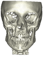
PAR-12-001: R01DE024450 University of Michigan Quantification Of 3d Bony Changes In Temporomandibular Joint Osteoarthritis The group at Michigan is working to precisely quantify three-dimensional bony changes in the Temporomandibular joint with the goal of improved early diagnosis and better monitoring of treatment outcomes. Funding Duration: 09/10/2013-08/31/2017 |
 
|
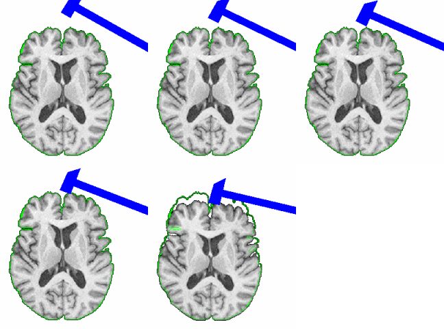
PAR-08-183: R21EB009900 Johns Hopkins Skull Stripping The group at Johns Hopkins is developing software that enables the stripping of skull, scalp, and meninges from structural MRI scans in a fully automated fashion. Funding Duration: 08/01/2009-07/31/2011 |
|
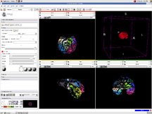
PAR-07-249: R01AA016748 Measuring Alcohol and Stress Interaction with Structural and Perfusion MRI This project is funded under an NCBC collaboration grant to PIs James Daunais, Robert Kraft, and Chris Wyatt. The goal of this project is to examine the the effects of chronic alcohol self-administration on brain structure and function the monkey brain. MRI image analysis tools from the NA-MIC kit will be adapted for use with the monkey brain datasets. More... Funding Duration: 07/15/2009-03/31/2010 |
|
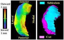
PAR-07-249: R01EB008171 3D Shape Analysis for Computational Anatomy This project is funded under an NCBC collaboration grant to PI Michael Miller JHU (with Joe Hennessey) Funding Duration: 05/21/2009-02/28/2013 |
|
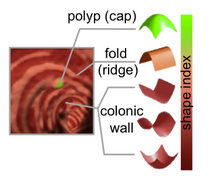
PAR-07-249: R01CA131718 NA-MIC Virtual Colonoscopy This project is funded under an NCBC collaboration grant to PI Hiroyuki Yoshida. The goal of this project is to More... Collaboration Duration: 01/26/2009-11/30/2009 |
|
PAR-07-249: R01MH084795 The Microstructural Basis of Abnormal Connectivity in Autism This project is funded under an NCBC collaboration grant to PI Janet Lainhart, MD. It will use tools developed within NAMIC for a longitudinal neuroimaging, clinical, and neuropsychological study of late neurodevelopment in autism.combining analysis of connectivity and morphometry. Funding Duration: 01/04/2009-01/31/2014 |
|
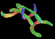
PAR-07-249: R01EB006733 Development and Dissemination of Robust Brain MRI Measurement Tools This project is funded under an NCBC collaboration grant to PI Dinggang Shen at UNC-Chapel Hill. The goal of this project is to develop and widely distribute a software package for robust measurement of brain structures in MR images using computational neuroanatomy methods.More... Funding Duration: 09/17/2008-08/31/2011 |
|
PAR-05-063: R01CA124377 An Integrated System for Image-Guided Radiofrequency Ablation of Liver Tumors This project is funded under an NCBC collaboration grant to PI Kevin Cleary at Georgetown University. The goal of this project is to develop and validate an integrated system based on open source software for improved visualization and probe placement during radiofrequency ablation (RFA) of liver tumors.More... Funding Duration: 09/14/2007-07/31/2012 |
|
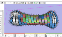
PAR-05-063: R01EB005973 Automated FE Mesh Development This project is funded under an NCBC collaboration grant to PIs Nicole Grosland and Vincent Magnotta at UIowa. The goal of this project is to integrate and expand methods to automate the development of specimen- / patient-specific finite element (FE) models into the NA-MIC kit. More... Funding Duration: 09/20/2006-06/30/2011 |
Additional External Collaborations
This section describes external collaborations with NA-MIC that are funded by other mechanisms:
|
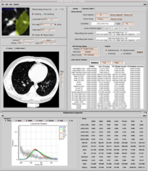
R01HL116931 Airway Inspector: a chest imaging biomarker software platform for COPD
Airway Inspector is a open source software tool based on Slicer 2.x developed at Brigham and Women’s Hospital for the analysis of chest CT scans for the analysis of emphysema and airway disease. The tool was conceived to analysis low resolution CT scans bringing the gap between retrospective data and new emerging CT technologies. The success of the tool in the COPD clinical community is supported by the array of peer-review publications that have used the tool to define metrics of disease that enable hypothesis driven research. Beside the existence of alternative commercial applications, Airway Inspector is the only freely available maintained platform for COPD research. In spite of its success, Airway Inspector is limited to a discontinued version of Slicer. The broad objective of this proposal is to support the refactoring and development of Airway Inspector as a platform for image-based COPD research. This goal will be achieved by the creation of the Chest Imaging Biomarker Platform (CIBP) library that integrates novel algorithm solutions to lung image analysis that have been developed in our laboratory. Those solutions include: robust lung extraction, parenchymal tissue classification based on local density, airway, fissure and pulmonary vascular extraction based on scale-space particles and airway labeling based on Hidden Markov Models. That software platform will be used to create workflows that will be integrated in Slicer 4 for their deployment in the clinical community. The workflows will provide an end-to-end solution for the clinical to obtain phenotypes to characterize emphysema, airway disease and pulmonary vascular remodeling. Pending Council Review Funding Duration: 12/1/2012 - 11/30/2017 |

|
R01HL116473 The clinical impact of pulmonary vascular remodeling in smokers
Smoking related pulmonary vascular disease has been demonstrated to be an independent predictor of morbidity and mortality in patients with COPD. Previous investigation and observation suggests that there are several types of pathologic pulmonary vascular remodeling possible in smokers. These range from inflammatory remodeling with progressive luminal occlusion, aberrant vessel elongation in regions of hyperinflation, and outright loss of vasculature in regions of severe emphysematous destruction. While there are existing tools that may be used to investigate the aggregate effect of these processes such as right heart catheterization, echocardiography, and measurements of diffusing capacity for carbon monoxide (DLCO), none can differentiate the types of vascular remodeling/deformation present nor their relative contribution to clinical impairment. The purpose of this investigation is to improve understanding of the clinical impact and epidemiologic associations of pulmonary vascular remodeling in smokers. We will do this by performing CT based quantitative measures of the mediastinal (Aim 1) and intra-parenchymal vasculature (Aim 2) in all subjects enrolled in the COPDGene Study and then clinically validating these measures with echocardiography, cardiac MRI and measures of DLCO (Aim 3). In the final steps of Aim 3 we will examine the overlap and associations between pulmonary and cardiovascular disease (both clinical diagnosed CVD and both coronary and thoracic aortic calcification) and the relationship between pulmonary vascular morphology and exercise capacity, acute exacerbations of COPD, symptoms, and mortality. We believe that this will lead to improved understanding of the pathophysiology of COPD and may ultimately improve the care of patients with COPD. Pending Council Review Funding Duration: 12/1/2012 - 11/30/2017 |
|
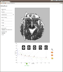
R01EB014947 MI2B2 ENABLED PEDIATRIC RADIOLOGICAL DECISION SUPPORT
This project is the direct successor to image informatics research supported by the mBIRN project. With the Medical Image Informatics Bench to Bedside system, extensive medical record databases can be linked to the corresponding medical image data in the institutional PACS. The pilot study funded here applies this technology to study of infant brain development by tapping into a large cohort of multi-parametric MR studies acquired during routine clinical care at Boston Children's Hospital. Funding Duration: 8/1/2012 - 7/31/2016 |

|
U24RR026057 Collaborative Tools Support Network for BIRN As the mBIRN and fBIRN test bed activities wind down the activities, research labs that have adopted the BIRN tool suite are supported through the BIRN-CTSN efforts. This funding covers software maintenance and ongoing deployment support to ensure that these important resources remain a vital part of the research community. Funding Duration: 09/30/2009-08/31/2013 |

|
U24RR025736 BIRN CC The NCRR-funded efforts of the BIRN Coordinating Center (BIRN-CC) strive to apply state-of-the-art computer science techniques to the growing problem of working with large biomedical informatics datasets. Through a series of test bed projects and use case studies, the BIRN-CC refines and optimizes its offerings. NA-MIC provides an opportunity for BIRN-CC to work closely with the image analysis community and learn from the experience of NA-MIC DBPs. Funding Duration: 12/15/2008-11/30/2013 |
|
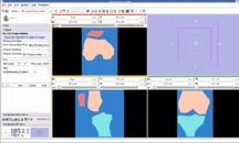
U54GM072970 NCBC Stanford Simbios Our sister NCBC at Stanford, dedicated to biomedical simulation, is working to adapt NA-MIC image analysis routines generate simulation models directly from MRI scans. Funding Duration: 09/19/2008-07/31/2010 |
| U54EB005149-04S1 NA-MIC Collaboration with NITRC The NA-MIC Project is working to make NA-MIC neuroimaging software available through the NITRC web site. Supplemental support is helping to create the Slicer3 Loadable Modules project so that slicer plugins can be hosted on NITRC, allowing greater scalability for developers and users of Slicer. Funding Duration: 08/01/2008-07/31/2009 | |

|
NAMIC supports COPDGene® quantitative analysis The Genetic Epidemiology of COPD (COPDGene®) Study is one of the largest studies ever to investigate the underlying genetic factors of Chronic Obstructive Pulmonary Disease or COPD. Through the enrollment of over 10,000 individuals, the COPDGene® Study aims to find inherited or genetic factors that make some people more likely than others to develop COPD. With the use of CT scans, COPDGene® also seeks to better classify COPD and understand how the disease may differ from person to person. Funding Duration: 09/27/2007-07/31/2012 |
|
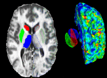
PAR-05-057: R01NS050568 BRAINS Morphology and Image Analysis This project is a funded under a Continued Development and Maintenance of Software grant to PIs Vincent Magnotta, Hans Johnson, Jeremy Bockholt, and Nancy Andreasen at the University of Iowa. The goal of this project is to update the BRAINS image analysis software developed at the University of Iowa. More... Funding Duration: 05/15/2007-03/31/2010 |
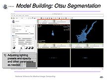
|
NCBC Supplement for Microscopy and Slicer An NCBC Supplement to NCMIR, UCSD focused on the utilization of Slicer with microscopy data and resulted in a tutorial for use of Slicer with confocal microscopy data. Funding Duration: 08/01/2006-07/31/2008 |
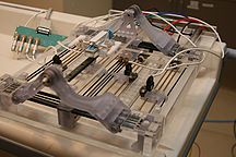
|
R01CA111288 NA-MIC Collaboration with Prostate BRP BRP is leveraging the NA-MIC kit as a platform for developing dedicated IGT capabilities. Funding Duration: 07/01/2006-05/31/2016 |
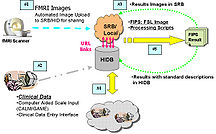
|
U24RR021992 fBIRN The Function BIRN (fBIRN) addresses the difficult problem of collecting and analyzing fMRI data collected at multiple sites in the context of schizophrenia research. A number of important and difficult scientific and engineering problems can only be addressed in the context of a multi-site consortium that aims to reproducibly quantify brain activity. Outcomes of fBIRN include quality assurance checks that have become industry standards, novel statistical approaches to identify and control for site-specific and scanner-specific biases, standardized stimulus paradigms, and fMRI informatics techniques for large scale datasets. Funding Duration: 02/08/2006-11/30/2010 |
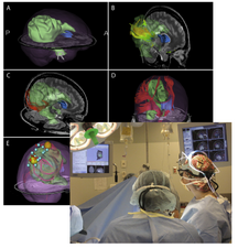
|
P41EB015898 NA-MIC Collaboration with NCIGT NCIGT is leveraging the NA-MIC kit as a platform for developing dedicated IGT capabilities. Funding Duration: 09/29/2005-07/31/2015 |
|
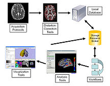
U54LM008748 NCBC I2B2 Our sister NCBC at Harvard Medical School, dedicated to biomedical image informatics, is working with us through the Harvard based, NCRR funded CTSC (Catalyst) program, to develop common open source software for the community. Funding Duration: 09/15/2004-7/31/2010 |
|
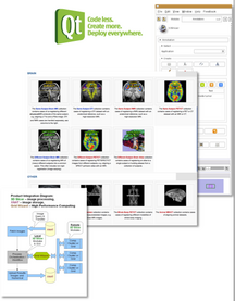
U24RR021382 mBIRN The Morphometry BIRN (mBIRN) seeks to support multi-site brain studies using structural and diffusion MRI. Standard acquisition protocols, analysis methods, and informatics techniques have all been studied and promulgated by the mBIRN community of researchers. Funding Duration: 09/30/2004-05/31/2010 |
|
P41RR013218 NA-MIC Collaboration with NAC NAC, the neuroimage analysis center, is a national resource center. NAC is relying on the NA-MIC kit for its general software environment. The mission of NAC is to develop novel concepts for the analysis of images of the brain and develop and disseminate tools based on those concepts. Several ARRA-funded supplements to the NAC grant have close ties to related efforts in NA-MIC. Funding Duration: 09/30/1998-05/31/2013 |
|
BrainColor brainCOLOR is a Collaborative Open Labeling Online Resource for to create high quality manually segmented brain data sets. |
|
Real-Time Computing for Image Guided Neurosurgery Using the Tera Grid to implement mesh-based non-rigid registration for Neurosurgery. |
|
UL1RR025758 NA-MIC support for Harvard CTSC Translational Imaging Consortium The Harvard CTSC Translational Imaging Consortium is using NA-MIC communication tools to facilitate the rapid deployment of expertise in medical imaging acquisition, analysis and visualization to clinical translational investigators. |
|
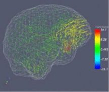
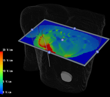
Children's Pediatric Cardiology Collaboration with SCI/SPL/Northeastern Collaboration with John Triedman, Matt Jolley, Dana Brooks, SCI. |
International Collaborations

|
Ontario Consortium of Adaptive Interventions for Radiation Oncology (OCAIRO) / Software Platform and Adaptive Radiotherapy Kit (SparKit) OCAIRO is a cross-Ontario initiative led by Dr. Jaffray, and will work towards developing adaptive radiation therapy--a new approach involving the creation of hardware, software, imaging and database systems to enable oncologists to adapt radiation to each individual patient and their response during the course of therapy. Cancer Care Ontario funded an additional project that focuses on developing a common Software Platform and Adaptive Radiotherapy Kit (SparKit) for OCAIRO and other cancer researchers in Ontario. |

|
Computer Aided and Image Guidance Medical Interventions (CO-ME) CO-ME The National Centre of Competence in Research (NCCR) Co-Me is a network of leading clinics and engineering sites in Switzerland with strong links to industry and international partners. |
|
NA-MIC Collaboration for Neurosurgical Intervention with University Hospital of Marburg Germany Marburg The neurosurgery Department of the University Hospital of Marburg is collaborating with NA-MIC, NCIGT, and NAC in medical image analysis and intervention technologies. |

|
Common Toolkit (CTK) CTK is a multi-institution international collaboration to share software development resources for medical imaging applications. |
|
Real Time Computer Simulation of Human Soft Organ Deformation for Computer Assisted Surgery |
| |
NA-MIC Collaboration with Research and Development Project on Intelligent Surgical Instruments Intelligent Surgical Instruments Projects uses Open-source software engineering tools developed by NA-MIC, and leverage it to surgical robotics. |
|
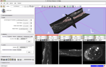
Vascular Modeling Toolkit Collaboration Slicer as a platform for segmentation and geometric analysis of vascular segments and image-based computational fluid dynamics (CFD). More... Collaboration with Luca Antiga of the Mario Negri Institute. |