Difference between revisions of "Projects:RegistrationLibrary:RegLib C19"
From NAMIC Wiki
(Created page with 'Back to ARRA main page <br> Back to Registration main page <br> [[Projects:RegistrationDocumentation:UseCaseInv…') |
|||
| Line 3: | Line 3: | ||
[[Projects:RegistrationDocumentation:UseCaseInventory|Back to Registration Use-case Inventory]] <br> | [[Projects:RegistrationDocumentation:UseCaseInventory|Back to Registration Use-case Inventory]] <br> | ||
| − | ==Slicer Registration Library Exampe #19: | + | ==Slicer Registration Library Exampe #19: Multi-contrast group analysis: intra- and inter-subject registration of multi-contrast MRI == |
{| style="color:#bbbbbb; background-color:#333333;" cellpadding="10" cellspacing="0" border="0" | {| style="color:#bbbbbb; background-color:#333333;" cellpadding="10" cellspacing="0" border="0" | ||
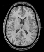
| − | |[[Image: | + | |[[Image:RegLib_C19_thumb_N2_T1.png|150px|this is the main reference image. All images are aligned into this space]] |
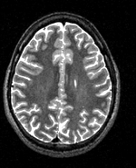
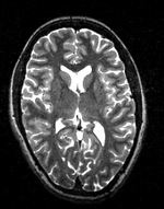
| − | |[[Image: | + | |[[Image:RegLib_C19_thumb_N2_T2.png|150px|this is the 1st intra-subject moving target, to be aligned with the main reference directly]] |
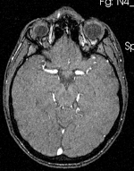
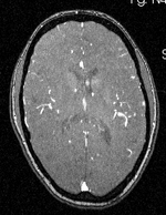
| + | |[[Image:RegLib_C19_thumb_N2_MRA.png|150px|this is the 2nd intra-subject moving target, to be aligned with the main reference directly]] | ||
|[[Image:Arrow_left_gray.jpg|100px|lleft]] | |[[Image:Arrow_left_gray.jpg|100px|lleft]] | ||
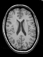
| − | |[[Image: | + | |[[Image:RegLib_C19_thumb_N4_T1.png|150px|this is both the fixed target for the 2nd subject and the moving image for inter-subject registration]] |
| − | |[[Image: | + | |[[Image:RegLib_C19_thumb_N4_T2.png|150px|this is a moving image to be aligned indirectly to the main ref]] |
| + | |[[Image:RegLib_C19_thumb_N4_MRA.png|150px|his is a moving image to be aligned indirectly to the main ref ]] | ||
|align="left"|LEGEND<br><small><small> | |align="left"|LEGEND<br><small><small> | ||
[[Image:Button_red_fixed.jpg|20px|lleft]] this indicates the reference image that is fixed and does not move. All other images are aligned into this space and resolution<br> | [[Image:Button_red_fixed.jpg|20px|lleft]] this indicates the reference image that is fixed and does not move. All other images are aligned into this space and resolution<br> | ||
[[Image:Button_green_moving.jpg|20px|lleft]] this indicates the moving image that determines the registration transform. <br> | [[Image:Button_green_moving.jpg|20px|lleft]] this indicates the moving image that determines the registration transform. <br> | ||
| − | |||
</small></small> | </small></small> | ||
|- | |- | ||
| − | |[[Image:Button_red_fixed.jpg|40px|lleft]] | + | |[[Image:Button_red_fixed.jpg|40px|lleft]] reference T1, subject 1 |
| − | |[[Image: | + | |[[Image:Button_green_moving.jpg|40px|lleft]] moving T2, subject 1 |
| + | |[[Image:Button_green_moving.jpg|40px|lleft]] moving MRA, subject 1 | ||
| | | | ||
| − | |[[Image: | + | |[[Image:Button_green_moving.jpg|40px|lleft]][[Image:Button_red_fixed.jpg|40px|lleft]] reference T1, subject 2 |
| − | |[[Image:Button_green_moving.jpg|40px|lleft]] | + | |[[Image:Button_green_moving.jpg|40px|lleft]] moving T2, subject 2 |
| + | |[[Image:Button_green_moving.jpg|40px|lleft]] moving MRA, subject 2 | ||
|- | |- | ||
| + | |1mm isotropic<br> 256 x 256 x 256<br>PA | ||
|1mm isotropic<br> 256 x 256 x 256<br>PA | |1mm isotropic<br> 256 x 256 x 256<br>PA | ||
|1mm isotropic<br> 256 x 256 x 256<br>PA | |1mm isotropic<br> 256 x 256 x 256<br>PA | ||
| | | | ||
| + | |0.9375 x 0.9375 x 1.5mm isotropic<br> 256 x 256 x 159<br>PA | ||
|0.9375 x 0.9375 x 1.5mm isotropic<br> 256 x 256 x 159<br>PA | |0.9375 x 0.9375 x 1.5mm isotropic<br> 256 x 256 x 159<br>PA | ||
|0.9375 x 0.9375 x 1.5mm isotropic<br> 256 x 256 x 159<br>PA | |0.9375 x 0.9375 x 1.5mm isotropic<br> 256 x 256 x 159<br>PA | ||
| Line 34: | Line 39: | ||
=== Keywords === | === Keywords === | ||
| − | MRI, brain, head, inter-subject, | + | multi-stage registration, MRI, brain, head, multi-contrast, inter-subject, group analysis, MRA, T2 |
===Input Data=== | ===Input Data=== | ||
| Line 46: | Line 51: | ||
#Build label mask of thalamus for A0: '''Editor''' module | #Build label mask of thalamus for A0: '''Editor''' module | ||
##Create Labelmap From”: ''A0_labels'' | ##Create Labelmap From”: ''A0_labels'' | ||
| − | |||
| − | |||
| − | |||
| − | |||
| − | |||
| − | |||
| − | |||
| − | |||
| − | |||
| − | |||
| − | |||
| − | |||
| − | |||
| − | |||
| − | |||
| − | |||
| − | |||
| − | |||
| − | |||
| − | |||
| − | |||
| − | |||
| − | |||
| − | |||
| − | |||
| − | |||
| − | |||
| − | |||
| − | |||
| − | |||
| − | |||
| − | |||
| − | |||
| − | |||
| − | |||
| − | |||
| − | |||
| − | |||
| − | |||
| − | |||
| − | |||
| − | |||
| − | |||
| − | |||
| − | |||
| − | |||
| − | |||
| − | |||
| − | |||
| − | |||
| − | |||
| − | |||
| − | |||
| − | |||
| − | |||
| − | |||
| − | |||
| − | |||
| − | |||
| − | |||
| − | |||
| − | |||
| − | |||
| − | |||
| − | |||
| − | |||
| − | |||
| − | |||
| − | |||
=== Registration Results=== | === Registration Results=== | ||
{| style="color:#bbbbbb; background-color:#333333;" cellpadding="10" cellspacing="0" border="0" | {| style="color:#bbbbbb; background-color:#333333;" cellpadding="10" cellspacing="0" border="0" | ||
| − | |[[Image: | + | |[[Image:RegLib_C19_unreg.gif|200px|left|unregistered]] [[Image:RegLib_C19_reg.gif|200px|left|registered]] |
|} | |} | ||
Revision as of 14:29, 2 July 2010
Home < Projects:RegistrationLibrary:RegLib C19Back to ARRA main page
Back to Registration main page
Back to Registration Use-case Inventory
Slicer Registration Library Exampe #19: Multi-contrast group analysis: intra- and inter-subject registration of multi-contrast MRI
Objective / Background
This is an example of inter-subject registration via surface matching. The structures of interest are a small subset of the entire image, hence registration is not driven by image intensities but rather two model surfaces derived from the labelmaps.
Keywords
multi-stage registration, MRI, brain, head, multi-contrast, inter-subject, group analysis, MRA, T2
Input Data
 reference/fixed : T1w coronal, 1mm isotropic. Called A1_gray
 reference/fixed : labelmap , aligned with above. Called A1_label
 moving: T1w coronal, 0.9 inplane, 1.5mm coronal slices. Called A0_gray
 moving: labelmap , aligned with above. Called A0_label
Methods
- Visualize & browse A0 data: determine label range of thalamic nuclei labels in A0_label: 500-526
- Visualize & browse A1 data: determine label range of thalamus lables in A1_label: 10 and 49
- Build label mask of thalamus for A0: Editor module
- Create Labelmap From”: A0_labels
Registration Results
Download
- download entire tutorial package (Original Data, Intermediate Results, Solution, zip file 74 MB)
- download old atlas dataset (MRI+labelmap,thalamic nuclei models, list of labels+names), zip file 5.4 MB)
- download new atlas dataset (MRI T1+T2, labelmap, thalamus model), zip file 126MB)
- download result merged new atlas dataset (MRI T1+T2, new merged labelmaps (2x) incl. thalamic nuclei, colormap file with label names), zip file 126 MB)
- download full step-by-step tutorial (PowerPoint, zip file, 2.5 MB)
- obsolete: download compare set (New Atlas, 2 merged versions (full & clipped), thalamus models, Xform, Merged colormap, zip file, 67 MB)
Link to User Guide: How to Load/Save Registration Parameter Presets
Discussion: Registration Challenges
- Because the structures of interest are a very small subset of the image without distinct grayscale contrast
- the two atlases represent different anatomies and hence some residual misalignment is inevitable
- the two labelmaps have different resolutions and different smoothness of structure outlines. Some need filtering to remove spurious surface details that would distract the registration algorithm
Discussion: Key Strategies
- Because the structures of interest are a very small subset of the image without distinct grayscale contrast, we co-register surfaces rather than intensity volumes
Acknowledgments
- dataset provided by Ron Kikinis, M.D. and Florin Talos, M.D.
