Difference between revisions of "Projects:RegistrationLibrary:RegLib C04"
| Line 6: | Line 6: | ||
=== Input === | === Input === | ||
{| style="color:#bbbbbb; " cellpadding="10" cellspacing="0" border="0" | {| style="color:#bbbbbb; " cellpadding="10" cellspacing="0" border="0" | ||
| − | |[[Image:Reglib_C04_Thumb_PD1.jpg| | + | |[[Image:Reglib_C04_Thumb_PD1.jpg|100px|lleft|this is the main fixed reference image. All images are ev. aligned into this space]] |
| − | |[[Image:Reglib_C04_Thumb_T21.jpg| | + | |[[Image:Reglib_C04_Thumb_T21.jpg|100px|lleft|this is the main fixed reference image. All images are ev. aligned into this space]] |
| − | |[[Image:RegArrow_Affine.png| | + | |[[Image:RegArrow_Affine.png|70px|lleft]] |
| − | |[[Image:Reglib_C04_Thumb_Gd1.jpg| | + | |[[Image:Reglib_C04_Thumb_Gd1.jpg|100px|lleft|this is the intra-subject moving image. ]] |
|- | |- | ||
|exam 1: PD | |exam 1: PD | ||
| Line 16: | Line 16: | ||
|exam 1: T1-Gd | |exam 1: T1-Gd | ||
|- | |- | ||
| − | |[[Image:RegArrow_AffineVert.png| | + | |[[Image:RegArrow_AffineVert.png|70px|lleft]] |
| | | | ||
| | | | ||
| | | | ||
|- | |- | ||
| − | |[[Image:Reglib_C04_Thumb_PD2.jpg| | + | |[[Image:Reglib_C04_Thumb_PD2.jpg|100px|lleft|this is the inter-subject moving image, but also the reference for exam 2]] |
| − | |[[Image:Reglib_C04_Thumb_T22.jpg| | + | |[[Image:Reglib_C04_Thumb_T22.jpg|100px|lleft|this is the inter-subject moving image, but also the reference for exam 2]] |
|[[Image:RegArrow_Affine.png|100px|lleft]] | |[[Image:RegArrow_Affine.png|100px|lleft]] | ||
| − | |[[Image:Reglib_C04_Thumb_Gd2.jpg| | + | |[[Image:Reglib_C04_Thumb_Gd2.jpg|100px|lleft|this is the moving image. ]] |
|- | |- | ||
|exam 2: PD | |exam 2: PD | ||
| Line 68: | Line 68: | ||
*we then nest the first alignment within the second | *we then nest the first alignment within the second | ||
*because of the contrast differences and anisotropic resolution we use Mutual Information as cost function for better robustness | *because of the contrast differences and anisotropic resolution we use Mutual Information as cost function for better robustness | ||
| − | + | === Procedure === | |
| + | #Load example dataset via OpenScene... | ||
| + | #Align Exam 1: | ||
| + | ##Open ''BrainsFit'' module | ||
| + | ##Fixed Image: e1_PD, moving image: e1_T1 | ||
| + | ## | ||
=== Registration Results=== | === Registration Results=== | ||
[[Image:RegLib_C04_T1Gd-PD_unreg_AnimGif.gif||500px|Unregistered baseline data: PD vs. T1Gd]] Unregistered baseline data: PD vs. T1Gd<br> | [[Image:RegLib_C04_T1Gd-PD_unreg_AnimGif.gif||500px|Unregistered baseline data: PD vs. T1Gd]] Unregistered baseline data: PD vs. T1Gd<br> | ||
Revision as of 14:40, 13 September 2010
Home < Projects:RegistrationLibrary:RegLib C04Back to ARRA main page
Back to Registration main page
Back to Registration Use-case Inventory
v3.6.1  Slicer Registration Library Case 04:
Intra-subject Brain MR of Multiple Sclerosis: Multi-contrast series for lesion change assessment
Input
|
|

|
|
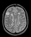
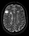
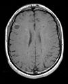
| exam 1: PD | exam 1: T2 | exam 1: T1-Gd | |

|
|||
|
|
|
|
| exam 2: PD | exam 2: T2 | exam 2: T1-Gd |
Modules
- Slicer 3.6.1 recommended modules: BrainsFit, Robust Multiresolution Affine
Objective / Background
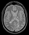
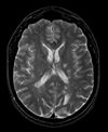
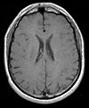
This scenario occurs in many forms whenever we wish to assess change in a series of multi-contrast MRI. The follow-up scan(s) are to be aligned with the baseline, but also the different series within each exam need to be co-registered, since the subject may have moved between acquisitions. Hence we have a set of nested registrations. This particular exam features a dual echo scan (PD/T2), where the two structural scans are aligned by default. The post-contrast T1-GdDTPA scan however is not necessarily aligned with the dual echo. Also the post-contrast scan is taken with a clipped field of view (FOV) and a lower axial resolution, with 4mm slices and a 1mm gap (which we treat here as a de facto 5mm slice).
Download
- Registration Library Case 04: MS Multi-contrast series (PD,T2, T1-GdDTPA): Lesion change assessment (Data,Presets, Solution, zip file 17 MB)
- download Registration Presets (BrainsFit) (mrml file ,12kB)
- download tutorial
Link to User Guide: How to Load/Save Registration Parameter Presets
Keywords
MRI, brain, head, intra-subject, multiple sclerosis, MS, multi-contrast, change assessment, dual echo, nested registration
Input Data
- reference/fixed : PD.1 baseline exam , 0.9375 x 0.9375 x 3 mm voxel size, axial acquisition, RAS orientation.
- fixed T2.1 baseline exam , 0.9375 x 0.9375 x 3 mm voxel size, axial acquisition, RAS orientation. -> (aligned with PD.1, not used for registering)
- moving: T1.1 (GdDTPA contrast-enhanced scan) baseline exam 0.9375 x 0.9375 x 5 mm voxel size, axial acquisition.
- moving: PD.2 follow-up exam 0.9375 x 0.9375 x 3 mm voxel size, axial acquisition.
- moving: T2.2 follow-up exam 0.9375 x 0.9375 x 3 mm voxel size, axial acquisition. -> same orientation as PD2, will have same transform applied
- moving:T1.2-GdDTPA follow-up exam0.9375 x 0.9375 x 5 mm voxel size, axial acquisition. -> undergoes 2 transforms: first to PD.2, then to PD.1
Registration Challenges
- we have multiple nested transforms: each exam is co-registered within itself, and then the exams are aligned to eachother
- potential pathology change can affect the registration
- anisotropic voxel size causes difficulty in rotational alignment
- clipped FOV and low tissue contrast of the post-contrast scan
Key Strategies
- we first register the post-contrast scans within each exam to the PD
- second we register the follow-up PD scan to the baseline PD
- we also move the T2 exam within the same Xform
- we then nest the first alignment within the second
- because of the contrast differences and anisotropic resolution we use Mutual Information as cost function for better robustness
Procedure
- Load example dataset via OpenScene...
- Align Exam 1:
- Open BrainsFit module
- Fixed Image: e1_PD, moving image: e1_T1
Registration Results
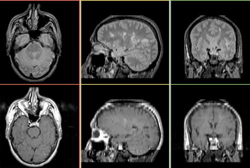 Unregistered baseline data: PD vs. T1Gd
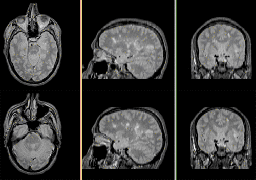 Unregistered followup data: PD exam 2 vs. exam 1
 Registered baseline data
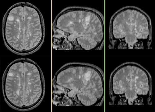 Registered followup data
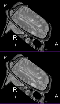 Lesion change visualization in 3D
 Lesion change via subtraction imaging of co-registered PD