Difference between revisions of "Projects:RegistrationLibrary:RegLib C10"
From NAMIC Wiki
| Line 32: | Line 32: | ||
*[[Image:Button_green_moving_white.jpg|20px]] moving: Probabilistic Tissue atlas, | *[[Image:Button_green_moving_white.jpg|20px]] moving: Probabilistic Tissue atlas, | ||
x 36 x 9 | x 36 x 9 | ||
| + | === Methods === | ||
| + | #build brain mask for fixed image using '''Skull Stripping''' module. Settings: 100 iterations, 20 subdivisions. New Volume: ''RegLib_C10_MRI_AtlasSegmentation_fixed_mask'' | ||
| + | #manually edit brain mask with Editor. required manual fix at frontal and occipital lobe | ||
| + | #run '''Register Images''' , Settings: | ||
| + | ::*Fixed Image: | ||
| + | ::*Moving Image: | ||
| + | ::*Resample Image: | ||
| + | ::*Load Transform: | ||
| + | ::*Save Transform: ''RegLib_C10_MRI_AtlasSegmentation_Xform_Affine_wmsk'' | ||
| + | ::*Initialization: Centers of Mass, | ||
| + | ::*Registration: PipelineAffine | ||
| + | ::*Expected offset: 10 | ||
| + | ::*Expected Rotation: 0.2 | ||
| + | ::*Expected Scale: 0.1 | ||
| + | ::*Expected Skew: 0.05 | ||
| + | ::*Fixed Image Mask: ''RegLib_C10_MRI_AtlasSegmentation_fixed_mask'' | ||
| + | ::*Affine Max Iteration: 80 | ||
| + | ::*Affine Sampling Ratio: 0.05 | ||
| + | ##(alternatively automated Affine Registration: '''Register Images Multires''' (Slicer 3.5) also produces good results | ||
| + | # run '"Deformable B-spline Registration'" module. Settings: | ||
| + | ::*Grid Size: 5 | ||
| + | :;*Histogram Bins: 50, | ||
| + | ::*Spatial Samples: 50000, | ||
| + | ::*initial transform: ''RegLib_C10_MRI_AtlasSegmentation_Xform_Affine_wmsk'' | ||
| + | |||
| + | |||
| + | |||
| + | |||
=== Registration Results=== | === Registration Results=== | ||
Revision as of 15:31, 17 February 2010
Home < Projects:RegistrationLibrary:RegLib C10Back to ARRA main page
Back to Registration main page
Back to Registration Use-case Inventory
Slicer Registration Library Exampe #10: Co-registration of probabilistic tissue atlas for subsequent EM segmentation
Objective / Background
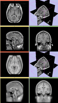
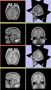
This is an example of sparse atlas co-registration. Not all atlases have an associated reference image that can be used for registration. Because the atlas represents a map of a particular tissue class probability, its contrast differs significantly from the target image.
Keywords
MRI, brain, head, inter-subject, probabilistic atlas, atlas-based segmentation
Input Data
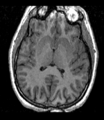
 reference/fixed : T1w axial, 1mm resolution in plane, 3mm slices
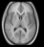
 moving: Probabilistic Tissue atlas,
x 36 x 9
Methods
- build brain mask for fixed image using Skull Stripping module. Settings: 100 iterations, 20 subdivisions. New Volume: RegLib_C10_MRI_AtlasSegmentation_fixed_mask
- manually edit brain mask with Editor. required manual fix at frontal and occipital lobe
- run Register Images , Settings:
- Fixed Image:
- Moving Image:
- Resample Image:
- Load Transform:
- Save Transform: RegLib_C10_MRI_AtlasSegmentation_Xform_Affine_wmsk
- Initialization: Centers of Mass,
- Registration: PipelineAffine
- Expected offset: 10
- Expected Rotation: 0.2
- Expected Scale: 0.1
- Expected Skew: 0.05
- Fixed Image Mask: RegLib_C10_MRI_AtlasSegmentation_fixed_mask
- Affine Max Iteration: 80
- Affine Sampling Ratio: 0.05
- (alternatively automated Affine Registration: Register Images Multires (Slicer 3.5) also produces good results
- run '"Deformable B-spline Registration'" module. Settings:
- Grid Size: 5
- Histogram Bins: 50,
- Spatial Samples: 50000,
- initial transform: RegLib_C10_MRI_AtlasSegmentation_Xform_Affine_wmsk
Registration Results
Download
- download entire package (Data,Tutorial, Solution, zip file xx MB)
- Presets
- Tutorial only
- Image Data only
Discussion: Registration Challenges
- Because the atlas represents a map of a particular tissue class probability, its contrast differs significantly from the target image.
- The two images may have strong differences in voxel sizes and voxel anisotropy. If the orientation of the highest resolution is not the same in both images, finding a good match can be difficult.
- The two images represent different anatomies, a non-rigid registration is required
Discussion: Key Strategies
- Because of the strong differences in image contrast, Mutual Information is recommended as the most robust metric.
- masking (skull stripping) is highly recommended to obtain good results.
- because speed is not that critical, we increase the sampling rate from the default 2% to 15%.
- we also expect larger differences in scale & distortion than with regular structural scans: so we significantly (2x-3x) increase the expected values for scale and skew from the defaults.
- a good affine alignment is important before proceeding to non-rigid alignment to further correct for distortions.


