Projects:RegistrationLibrary:RegLib C02
From NAMIC Wiki
Revision as of 15:22, 28 May 2010 by Meier (talk | contribs) (moved Projects:RegistrationDocumentation:RegLib C02 MultipleSclerosis to Projects:RegistrationLibrary:RegLib C02: making naming scheme consistent & suitable for automation)
Home < Projects:RegistrationLibrary:RegLib C02
Back to ARRA main page
Back to Registration main page
Back to Registration Use-case Inventory
Contents
Slicer Registration Use Case Exampe: Intra-subject Brain MR FLAIR to MR T1
Objective / Background
This scenario occurs in many forms whenever we wish to align all the series from a single MRI exam/session into a common space. Alignment is necessary because the subject likely has moved in between series.
Keywords
MRI, brain, head, intra-subject, FLAIR, T1, defacing, masking, labelmap, segmentation
Input Data
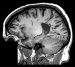
 reference/fixed : T1 SPGR , 1x1x1 mm voxel size, sagittal, RAS orientation.
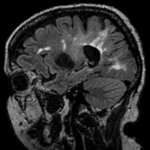
 moving: T2 FLAIR 1.2x1.2x1.2 mm voxel size, sagittal, RAS orientation.
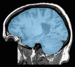
 mask: skull stripping labelmap obtained from SPGR, serves as mask
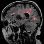
 tag: segmentation labelmap obtained from FLAIR.
- Content preview: Have a quick look before downloading: Does your data look like this? SPGR Lighbox , FLAIR Lighbox
Registration Challenges
- the amount of misalignment to be small. Subject did not leave the scanner in between the two acquisitions, but we have some head movement.
- we know the underlying structure/anatomy did not change, but the two distinct acquisition types may contain different amounts of distortion
- the T1 high-resolution had a "defacing" applied, i.e. part of the image containing facial features was removed to ensure anonymity. The FLAIR is lower resolution and contrast and did not need this. The sharp edges and missing information in part of the image may cause problems.
- we have a skull stripping label map of the fixed image (T1) that we can use to mask out the non-brain part of the image and prevent it from actively participating in the registration.
- we have one or more label-maps attached to the moving image that we also want to align.
- the different series have different dimensions, voxel size and field of view. Hence the choice of which image to choose as the reference becomes important. The additional image data present in one image but not the other may distract the algorithm and require masking.
- hi-resolution datasets may have defacing applied to one or both sets, and the defacing-masks may not be available
- the different series have different contrast. The T1 contains good contrast between white (WM) and gray matter (GM) , and pathology appears as hypointense. The FLAIR on the other hand shows barely any WM/GM contrast and the pathology appears very dominantly as hyperintense.
Key Strategies
- we choose the SPGR as the anatomical reference. Unless there are overriding reasons, always use the highest resolution image as your fixed/reference, to avoid loosing data through the registration.
- the defacing of the SPGR image introduces sharp edges that can be detrimental. We apply a multiresolution scheme at least. If this fails we mask that area or better still the brain. As a general rulle, if you have the mask available, use it.
- because of the contrast differences and the defacing we use Mutual Information as the cost function.
- because of the combined effects of rotational misalignment, defacing, pathology and contrast differences, we use a multi-resolution approach (Register Images MultiRes).
Registration Results
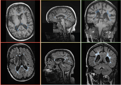 Registration Result: FLAIR + segmentation aligned with SPGR
Download
- Registration Library Case 02: MSBrain intra-subject multi-contrast: FLAIR+lesion segmentation to SPGR + mask (Data,Presets, Solution, zip file 27 MB)
- download/view guided video tutorial
- download power point tutorial
- download step-by step text instructions (text only)
Link to User Guide: How to Load/Save Registration Parameter Presets
