NA-MIC VMTK Collaboration
From NAMIC Wiki
Home < NA-MIC VMTK Collaboration
Collaboration with Luca Antiga of the Mario Negri Institute.
Back to NA-MIC_External_Collaborations
Goals of the Project
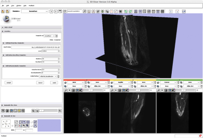
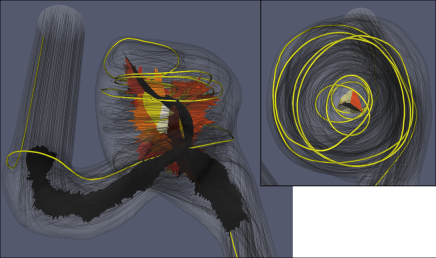
To create a Slicer-based platform for segmentation of vascular segments from angiographic images (CTA, MRA, ...), analysis of vascular geometry, mesh generation and hemodynamic simulation (CFD) and visualization.
Since many of these functionalities are already provided by the Vascular Modeling Toolkit (www.vmtk.org) as command-line tools, the plan is to integrate vmtk scripts within Slicer as command-line modules for the non-interactive tasks, and to create ad-hoc GUI modules for the more interactive tasks (i.e. segmentation).
Current progress
- vmtk script for automated conversion of vmtk command line pipes to Slicer command-line modules (DONE)
- Implementation of vmtk-based interactive modules (IN PROGRESS)
References
2007 Summer Project Week 2008 Summer Project Week 2009 Winter Project Week
Citations
- Lee SW, Antiga L, Spence JD and Steinman DA. Geometry of the carotid bifurcation predicts its exposure to disturbed flow. Stroke. Accepted.
- Thomas JB, Antiga L, Che S, Milner JS, Hangan Steinman DA, Spence JD, Rutt BK and Steinman DA. Variation in the carotid bifurcation geometry of young vs. older adults: Implications for "geometric risk" of atherosclerosis. Stroke, 36(11): 2450-2456, Nov 2005.
- Antiga L, Steinman DA. Robust and objective decomposition and mapping of bifurcating vessels. IEEE Transactions on Medical Imaging, 23(6): 704-713, June 2004.
- Antiga L, Ene-Iordache B and Remuzzi A. Computational geometry for patient-specific reconstruction and meshing of blood vessels from MR and CT angiography. IEEE Transactions on Medical Imaging, 22(5): 674-684, May 2003.

