Projects:RegistrationLibrary:RegLib C04
Back to ARRA main page
Back to Registration main page
Back to Registration Use-case Inventory
v3.6.1  Slicer Registration Library Case 04:
Intra-subject Brain MR of Multiple Sclerosis: Multi-contrast series for lesion change assessment
Input
|
|
|
|

|
|||
|
|

|
|
Modules
- Slicer 3.6.1 recommended modules: BrainsFit, Robust Multiresolution Affine
| |
| |
| |
|![]() |
| |
| |
| |align="left"|LEGEND
![]() this indicates the reference image that is fixed and does not move. All other images are aligned into this space and resolution
this indicates the reference image that is fixed and does not move. All other images are aligned into this space and resolution
![]() this indicates the moving image that determines the registration transform.
this indicates the moving image that determines the registration transform.
![]() this indicates images that passively move into the reference space, i.e. they have the transform applied but do not contribute to the calculation of the |0.9375 x 0.9375 x 3 mm axial
this indicates images that passively move into the reference space, i.e. they have the transform applied but do not contribute to the calculation of the |0.9375 x 0.9375 x 3 mm axial
256 x 256 x 51
RAS
|0.9375 x 0.9375 x 3 mm axial
256 x 256 x 51
RAS
|0.9375 x 0.9375 x 5 mm axial
256 x 256 x 24
RAS
|
|0.9375 x 0.9375 x 3 mm axial
256 x 256 x 54
RAS
|0.9375 x 0.9375 x 3 mm axial
256 x 256 x 54
RAS
|0.9375 x 0.9375 x 5 mm axial
256 x 256 x 24
RAS
Objective / Background
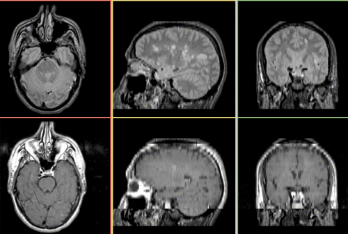
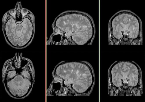
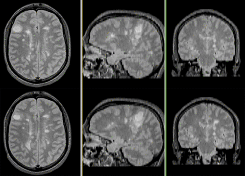
This scenario occurs in many forms whenever we wish to assess change in a series of multi-contrast MRI. The follow-up scan(s) are to be aligned with the baseline, but also the different series within each exam need to be co-registered, since the subject may have moved between acquisitions. Hence we have a set of nested registrations. This particular exam features a dual echo scan (PD/T2), where the two structural scans are aligned by default. The post-contrast T1-GdDTPA scan however is not necessarily aligned with the dual echo. Also the post-contrast scan is taken with a clipped field of view (FOV) and a lower axial resolution, with 4mm slices and a 1mm gap (which we treat here as a de facto 5mm slice).
Keywords
MRI, brain, head, intra-subject, multiple sclerosis, MS, multi-contrast, change assessment, dual echo, nested registration
Input Data
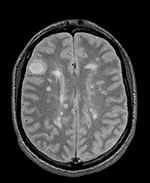
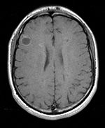
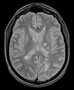
 reference/fixed : PD baseline exam , 0.9375 x 0.9375 x 3 mm voxel size, axial acquisition, RAS orientation.
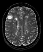
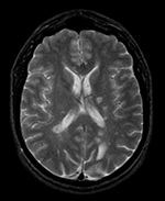
 reference/fixed : T2 baseline exam , 0.9375 x 0.9375 x 3 mm voxel size, axial acquisition, RAS orientation.
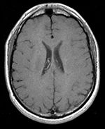
 moving: T1-GdDTPA baseline exam 0.9375 x 0.9375 x 5 mm voxel size, axial acquisition.
 moving: PD follow-up exam 0.9375 x 0.9375 x 3 mm voxel size, axial acquisition.
 tag: T2 follow-up exam 0.9375 x 0.9375 x 3 mm voxel size, axial acquisition.
 moving:T1-GdDTPA follow-up exam0.9375 x 0.9375 x 5 mm voxel size, axial acquisition.
Registration Challenges
- we have multiple nested transforms: each exam is co-registered within itself, and then the exams are aligned to eachother
- potential pathology change can affect the registration
- anisotropic voxel size causes difficulty in rotational alignment
- clipped FOV and low tissue contrast of the post-contrast scan
Key Strategies
- we first register the post-contrast scans within each exam to the PD
- we also move the T2 exam within the same Xform
- second we register the follow-up PD scan to the baseline PD
- we then nest the first alignment within the second
- we compare T1Gd to T1Gd, which went through 2 transforms. We correct residual misalignment with a 3rd correction transform
- because of the contrast differences and anisotropic resolution we use Mutual Information as cost function for better robustness
- we adjust the expected translation and rotational parameter settings to the visual assessment of the input data (i.e. initial misalignment)
Registration Results
Download
- Registration Library Case 04: MS Multi-contrast series (PD,T2, T1-GdDTPA): Lesion change assessment (Data,Presets, Solution, zip file 17 MB)
- download/view guided video tutorial
- download power point tutorial
Link to User Guide: How to Load/Save Registration Parameter Presets
