Events:RSNA 2010

|

|

|
|
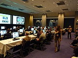
Slicer visualization tutorial
Slicer visualization tutorial
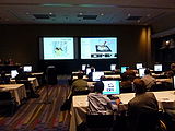
Slicer CTSC tutorial
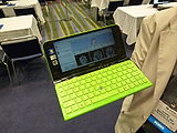
We ran out of RSNA computers, and one of the attendees downloaded and installed Slicer on his netbook!
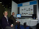
At the Slicer booth in Quantitative Imaging Reading Room
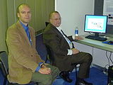
At the Slicer booth


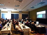
NA-MIC and NAC at RSNA
Introduction
The 3D Interactive Visualization of DICOM Images for Radiology Applications course is offered by the National Alliance for Medical Image Computing (NA-MIC) in conjunction with the Neuroimage Analysis Center (NAC) at the 96th Scientific Assembly and Annual Meeting of the Radiological Society of North America (RSNA 2010).
As part of the outreach missions of these NIH funded National Centers, we have developed an offering of freely available, multi-platforms open source software to enable medical image analysis research. The course along with the tutorial 3D Visualization datasets aim to introduce translational clinical scientists to the capabilities of the 3D Slicer software.
Logistics
- Date: Monday November 29, 2010
- Time: 2:30-4:00 pm
- Location: S401CD, McCormick Place, Chicago, Illinois
- Course: IA22
Learning Objectives
- Enhance interpretation of DICOM images through the use of computer-assisted 3D visualization,
- Enhance the understanding of the correlation of the segments of the liver with the surrounding vascular anatomy,
- Introduce cutting-edge open-source computer graphics applications for Radiology.
Abstract
Three-dimensional visualization of the anatomy is being made possible through the combined emergence of technological breakthroughs in Radiology hardware and increasingly sophisticated software tools for medical image analysis.
For the past six years, the National Alliance for Medical Image Computing (NA-MIC), one of the seven National Centers for Biomedical Computing, part of the NIH Roadmap for medical research, has converted some of the major scientific advances made by the biomedical imaging community into open-source software tools, contributing fasten the deployment of cutting-edge visualization techniques on a national and international scale.
As part of the NA-MIC toolkit, the 3D SLICER open-source software has been developed as a technology delivery platform for clinical researchers. 3D SLICER has become a multi-institution effort to share the latest advances in image analysis with the scientific and clinical community. To foster the translation of NA-MIC technology to clinical research applications, the NA-MIC Training Core has developed a series of workshop and tutorials using the 3DSLICER software.
This workshop is an introduction to the basics of viewing and interacting with DICOM volumes in 3D using the SLICER software. The 90 minute course is divided into two sections: the first part introduces the concepts of 3D visualization through an hands-on training session using two MR DICOM datasets of the brain and 3D reconstructed models of cerebral structures; the second part guides the user through the exploration of abdominal structures using a series of models, which include the segments of the liver, reconstructed from DICOM images of three clinical cases. Interactions with the 3D models are fostered by a series of ten radiological tasks to accomplish by the participants. Detailed answers to the tasks are provided during the workshop as the instructors guide the audience through the 3D visualization settings to enhance the understanding of the complexity of the anatomical structures involved.
Instructors
- Author/Presenter: Kitt Shaffer MD, PhD
- Author/Presenter: Sonia Marie-Aurore Pujol PhD
- Author/Presenter: Randy L. Gollub MD, PhD
- Andriy Fedorov, PhD
- Wendy Plesniak, PhD
- Ron Kikinis, MD
How to sign up
CTSA at RSNA
Introduction
The Quantitative Medical Imaging for Clinical Research and Practice course is offered by the Clinical Translational Science Award (CTSA) Consortium at the 96th Scientific Assembly and Annual Meeting of the Radiological Society of North America (RSNA 2010).
As part of the outreach missions of these cooperating institutions, we will offer a presentation and hands-on demonstration using freely available, multi-platforms open source software to enable medical image analysis research. The course along with the tutorial and datasets aim to introduce translational clinical scientists to the capabilities of the 3D Slicer software.
Logistics
- Date: Tuesday, November 30 2010
- Time: 10:30 AM - 12:00 PM
- Location: S401CD, McCormick Place, Chicago, Illinois
- Course: IA31
Learning Objectives
- Enhance interpretation of medical images through the use of 3D visualization,
- Gain experience with interactive, quantitative assessment of complex anatomical structures,
- Present current directions of quantitative imaging as a biomarker in clinical trials.
Abstract
Technological breakthroughs in medical imaging hardware and the emergence of increasingly sophisticated image processing software tools permit the visualization and display of complex anatomical structures with increasing sensitivity and specificity.
This workshop will begin with an introductory presentation of state-of-the-art, clinical examples of quantitative imaging biomarkers for diagnosis and clinical trial outcome measures. Cases from multiple imaging modalities and from multiple organ systems will be highlighted to illustrate the depth and breath of this field. Participants will then be led through a series of tutorials on the basics of viewing and processing DICOM volumes in 3D using 3D SLICER (www.SLICER.org). Specific hands-on demonstrations will focus on basic use of 3D Slicer software, quantitative measurements from PET/CT studies, and volumetric analysis of meningioma.
Instructors
- Author/Presenter: Randy L. Gollub MD, PhD
- Author/Presenter: Katarzyna J. Macura MD, PhD
- Author/Presenter: Jeffrey T. Yap PhD
- Author/Presenter: Ron Kikinis MD
- Author/Presenter: Valerie Humblet
- Author/Presenter: Wendy Plesniak PhD
Tutorial Materials
How to sign up
3D Slicer at RSNA
Quantitative Imaging Reading Room Exhibit
Title: 3D Slicer: An Open Source Application for Registration, Segmentation, Quantitative Analysis, and Visualization of Biomedical Image Data
- Focus: Interactive Editor, Volume Rendering, Tools for Quantitative Analysis
Exhibitors
- A Fedorov
- K. Hayes
- W. Plesniak
- R. Kikinis
- V. Humblet
"Meet the Experts" sessions for this exhibit will be held on the following dates/times:
- Monday November 29 (12:15-1:15pm)
- Wednesday December 1 (12:15-1:15pm)
- Thursday December 2 (12:15 - 1:15pm)
Logistics
The Quantitative Imaging Reading Room will be located in the Lakeside Learning Center (Hall E) of McCormick Place Convention Center, Chicago. The Lakeside Learning Center will be open according to the following schedule:
- Sunday, Nov. 28, 8:00am – 6:00pm
- Monday-Thursday, Nov. 29-Dec. 2, 7:00am – 10:00pm
- Friday, Dec. 3, 7:00am – 12:45pm
Back to NA-MIC Events