Home < Project Week 25 < Wrist Kinematics:Back to Projects List
Key Investigators
Project Description
| Objective
|
Approach and Plan
|
Progress and Next Steps
|
- The clinical goal of this project is the use of dynamic MRI (instead of static series of 3D CT done so far) to explore 3D carpal dynamics to detect kinematic wrist abnormalities in diagnosis/staging/management, to study carpal motion in health/disease, and to evaluate results after surgery
- Collaborators: Catherine N. Petchprapa, MD, NYU Langone Musculoskeletal Imaging & Radiology, and Riccardo Lattanzi, NYU Langone Radiology.
- Challenges for image analysis include segmentation of wrist bones from a longitudinal series of relatively low resolution MRI with anisotropic voxels, consistent segmentation of individual bones across the time series, and joint spatiotemporal modeling of extracted bones as a multi-object complex.
|
- We will use Slicer and various tools for visualization, segmentation, and analysis of the dynamic MR sequence.
|
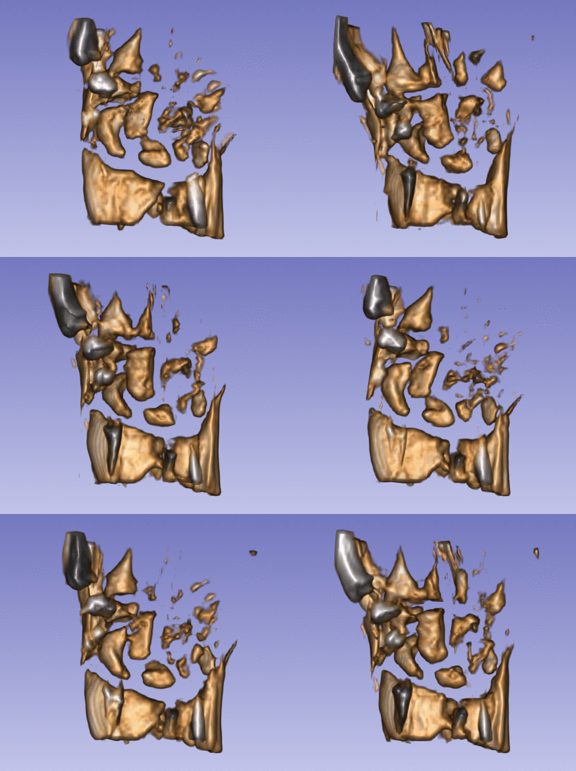
- Slicer was used to import the DICOM sequence and export 10 individual volumes. The sequences module combined with volume rendering within Slicer provided exploratory qualatative analysis of the wrist kinematics.
- The 10 images in the sequence were used to estimate a template image with ANTs.
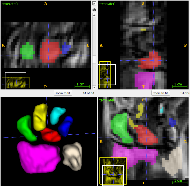
- 3D segmentation of individual bones was solved via level-set segmentation within itkSNAP. Preliminary tests with Slicer GrowCut were not satisfactory due to highly non-isotropic voxels, but more tests will be necessary to explore new advanced features within "Segment Editor".
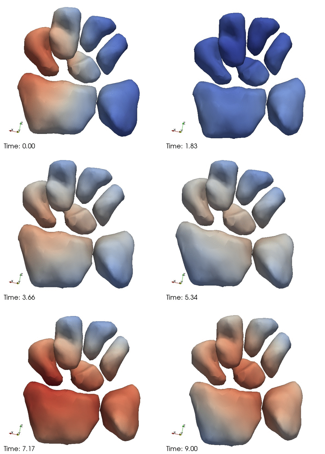
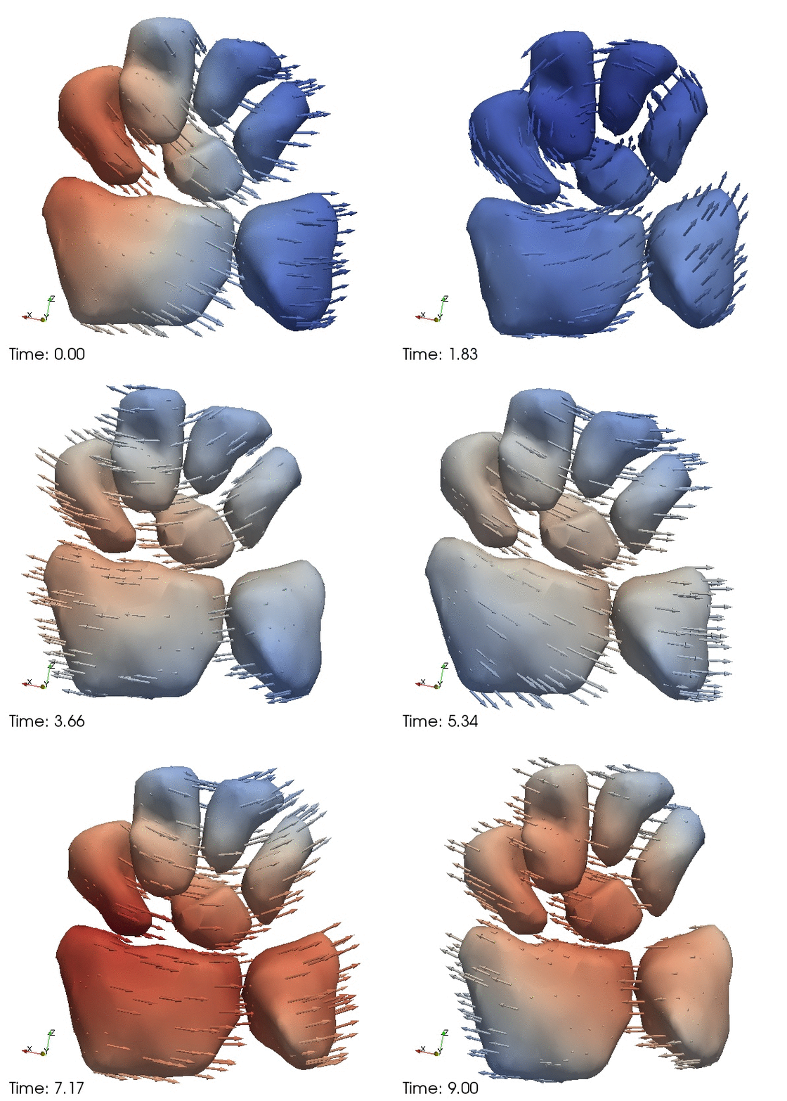
- Spatiotemporal modeling was performed jointly with all bones using shape4d.
|
Illustrations
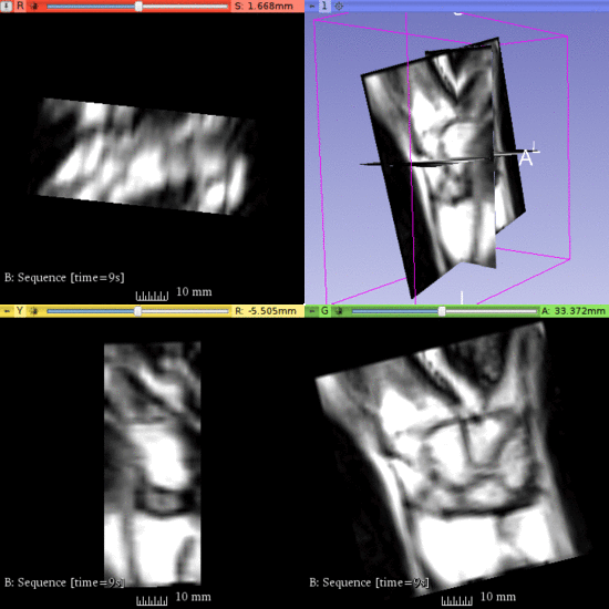
Left) One observation of the wrist. Right) A sequence of volume renderings within Slicer to show the 10 observations.
Segmentation of the wrist bones in the template space, which are propagated back to the individual observations via mappings computed during template estimation.
Continous change trajectories via spatiotemporal modeling. Color denotes speed and vectors denote direction of change.
Background and References
[1] Fishbaugh, J., Durrleman, S., Prastawa, M., Gerig, G. Geodesic shape regression with multiple geometries and sparse parameters. Medical Image Analysis. Vol 39. pp. 1-17. (2017)
[2] Fishbaugh, J., Durrleman, S., Gerig, G. Estimation of smooth growth trajectories with controlled acceleration from time series shape data. Proc. of Medical Image Computing and Computer Assisted Intervention (MICCAI '11). Vol 6892, pp. 401-408. (2011)
