Projects:LiverFibrosisStaging
Liver Fibrosis Staging
This project is conducted in collaboration with the Boston Medical Center. We are trying to provide tools for robust liver fibrosis staging, based on MRI image analysis. The current practice of fibrosis assessment, which is based on painful liver biopsy, might be dangerous. Moreover, the decision of the pathologist based on a biopsy is subjective, and depends on the sample, because the fibrosis level varies along the liver. No objective standard has been developed yet for histological fibrosis assessment. Magnetic resonance volume data has much lower resolution than histological image data, but it includes the entire liver volume. Also, MRI is non-invasive and not painful, thus it is preferred as a diagnostic tool. Previously it has been hypothesized that the average brightness of Apparent Diffusion Coefficient (ADC) in diffusion MRI correlates with the fibrosis stage.
Our Approach
In our research, we have tested different ADC image texture features, and have found features (e.g., standard deviation of color distribution) that correlate with histological results much better than average brightness. Using the tools from visual tracking research, we have developed algorithm for automatic hepatic MRI segmentation, and algorithm for automatic liver staging, based on optimal feature grouping. In addition, we are developing novel topological continuous features to improve the classification.
Results
The original liver slice for b=4 is shown in the Figure 1. The automatically segmented slice converted to Apparent Diffusion Coefficient (ADC) image, and shown in the Figure 2. Using multiple ADC image features we are able to provide automatic grading of hepatic fibrosis which correlate more than 80% with the histopathology results. This result improves the current state-of-the-art algorithms that able to classify livers only to two groups (healthy/unhealthy), with the similar correlation.
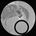
Figure 1. The original MRI data.

Figure 2. Automatically segmented MRI ADC.


Publications
Nakhmani A., Anderson. S., Tannenbaum A., "Automatic Segmentation and Classification of Hepatic Fibrosis Grade Using Diffusion MRI," In preparation.
Key Investigators
- UAB: Arie Nakhmani and Allen Tannenbaum