Semiautomatic longitudinal segmentation of MR volumes in traumatic brain injury
From NAMIC Wiki
Home < Semiautomatic longitudinal segmentation of MR volumes in traumatic brain injury
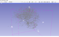
Fiber tract of TBI patient

Concept of visualization of deformation field and tissue change (from James Fishbaugh)


Editing Semiautomatic longitudinal segmentation and registration of MR volumes in traumatic brain injury
Key Investigators
- UCLA: Andrei Irimia, Micah Chambers, Jack Van Horn
- Kitware: Danielle Pace, Stephen Aylward
- University of Utah: Bo Wang, Marcel Prastawa, Guido Gerig
Objective
- For the purposes of this project, we have been interacting frequently for the purpose of developing methods for analyzing MR volumes of traumatic brain injury.
- The goal is to provide tools for the automatic segmentation of TBI from multimodal neuroimaging volume data.
- The Utah team has been focusing on the development of semi-automatic segmentation of TBI volumes, and the UCLA team has made appreciable progress on patient data analysis and quantification of results.
- At UCLA, we have acquired additional data sets of MRI/DTI from TBI patients. These data were shared with our colleagues in Utah and, at the meeting in Boston, we plan on working to investigate the performance of the Utah algorithm on these data sets.
- We have also been sharing ideas regarding how best to incorporate these tools into Slicer, and will continue to do so at the Meeting.
- For quantitatively analyzing the recovery and treatment efficacy of MR images with traumatic brain injury (TBI), we introduce a multimodal image segmentation framework for longitudinal TBI images.
- In the case of longitudinal images with TBI present topological changes over time due to the effect of the impact force and the recovery process, we propose a novel atlas construction scheme that explicitly models the effect of topological changes [1].
- A VTK development goal is to visualize graph and tract statistics using slicer
Approach
- The UCLA and Utah teams will be interacting and providing feedback with regard to an upcoming Slicer implementation of an automatic segmentation algorithm for TBI. This algorithm has been designed by Dr. Gerig's group and has been successfully applied to TBI data sets acquired at UCLA.
- During the Project Week we will focus on the analysis of additional subjects as well as on making plans to create a Slicer module to perform segmentation of TBI volumes via personalized atlas creation.
Additional approaches include:
- systematic inspection of existing TBI data and discussion on missing modalities and artifacts in some subject data
- discussion on how best to visualize TBI segmentation results to aid clinicians
- Creating a tool to load graph statistics into VTK polyline format so that properties of individual brain connectivity may be easily explored
Progress
- Andrei Irimia and Bo Wang participated in a breakout session on difficult registration, where ideas were exchanged with regard to various registration algorithms which other NA-MIC researchers have available for difficult data. One outcome of this meeting was the identification of various algorithms which can be used for TBI registration across time points. Other meeting participants included Stephen Aylward from Kitware, as well as Matt Toews and Sandy Wells from BWH. TBI data already available from UCLA were pointed out for other NA-MIC collaborators to explore and use with their registration methods.
- Bo Wang and Andrei Irimia performed a systematic inspection of available UCLA TBI data. Problematic data sets were discussed and some additional data were obtained from UCLA for one data set. Some data analysis was performed to determine how Slicer can best be used to extract clinically meaningful information from it.
- Novel algorithms from the Utah team which can be used in Slicer were identified and described. Bo and Andrei agreed to start working on two new TBI-related projects which may yield novel biomarkers of disease outcome. One involves the use of Slicer to visualize cortical atrophy in TBI using a personalized approach; the other makes use of Slicer to visualize changes in the shape of the ventricular system.
- Micah Chambers discussed with members of the slicer team the possibility of extending diffusion weighted images to extract connection matrices. In particular, he discussed the format of Slicer diffusion based tracts, and how our existing connectivity methods could be adapted to the slicer framework.
- Micah Chambers worked on graph theoretic metrics for calculating connectivity from VTK images.
Delivery Mechanism
We foresee that the Utah algorithm will be made available in Slicer upon further testing and evaluation. This work will be delivered to the NA-MIC Kit as a
- Slicer Module
- Slicer 4 Tutorial
References
[1] Bo Wang, Marcel Prastawa, Suyash P. Awate, Andrei Irimia, Micah C. Chambers, Paul M. Vespa, John D. Van Horn, Guido Gerig, Segmentation of Serial MRI of TBI patients using Personalized Atlas Construction and Topological Change Estimation, Proc. IEEE ISBI 2012, to appear May 2012, in print