File list
From NAMIC Wiki
This special page shows all uploaded files.
| Date | Name | Thumbnail | Size | Description | Versions |
|---|---|---|---|---|---|
| 13:45, 25 March 2011 | Sampling spatial-parameters.png (file) |  |
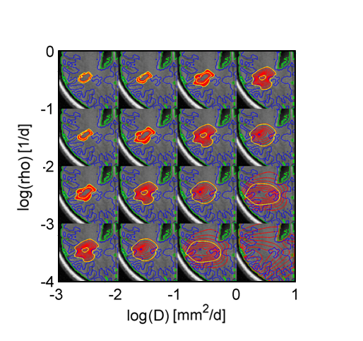
399 KB | Adaptive sampling of the parameter space for the synthetic high-grade data set. Sampling points (green) for the {x; y} coordinates of the initial tumor growth location. The figure also shows isolines of tumor cell density (red), the predicted extensions o | 1 |
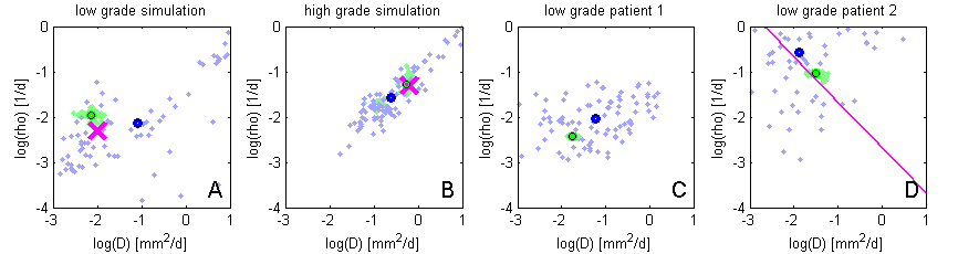
| 22:47, 24 March 2011 | Results Tumor-Model.png (file) | 15 KB | MCMC sampling results in the space spanned by model parameters D and rho, for the four experiments. Green samples are obtained from the sparse grid interpolation Eq. (12), blue-purple samples come from the direct sampling in Eq. (10). Black circles indica | 1 | |
| 21:32, 23 March 2011 | Fisher-state-space tumor-shapes.png (file) |  |
257 KB | Parameter space of a Fisher-type reaction-diffusion tumor model with tumor cell diffusivity D and proliferation rate rho. Shown are the shapes of tumors with the same size but different parametrization. | 1 |
| 12:23, 25 June 2010 | Mrsi slicer sivic.jpg (file) |  |
340 KB | 1 | |
| 12:17, 25 June 2010 | Mrsi slicer sivic.tif (file) | 413 KB | Reverted to version as of 12:13, 25 June 2010 | 4 | |
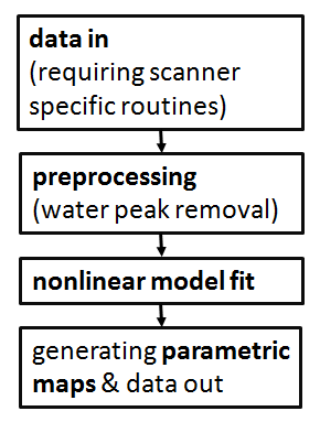
| 15:43, 8 January 2010 | Workflow MRSI signal processing.png (file) |  |
10 KB | 1 | |
| 18:26, 11 September 2009 | Tumor model.jpg (file) |  |
7 KB | 1 | |
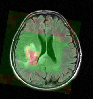
| 19:37, 10 September 2009 | Multimodal glioma.png (file) |  |
230 KB | 1 | |
| 19:30, 10 September 2009 | Tumor segmentation lesion atlas.png (file) |  |
96 KB | 1 | |
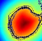
| 14:46, 25 June 2009 | Mrsi parametric-map-NAA 3d.jpg (file) |  |
100 KB | 2 | |
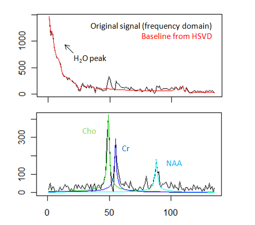
| 02:06, 5 December 2008 | Spectral fitting mrs.png (file) |  |
32 KB | Spectral fitting of magnetic resonance spectra. | 1 |
| 13:11, 9 June 2008 | Mrsi slicer.jpg (file) |  |
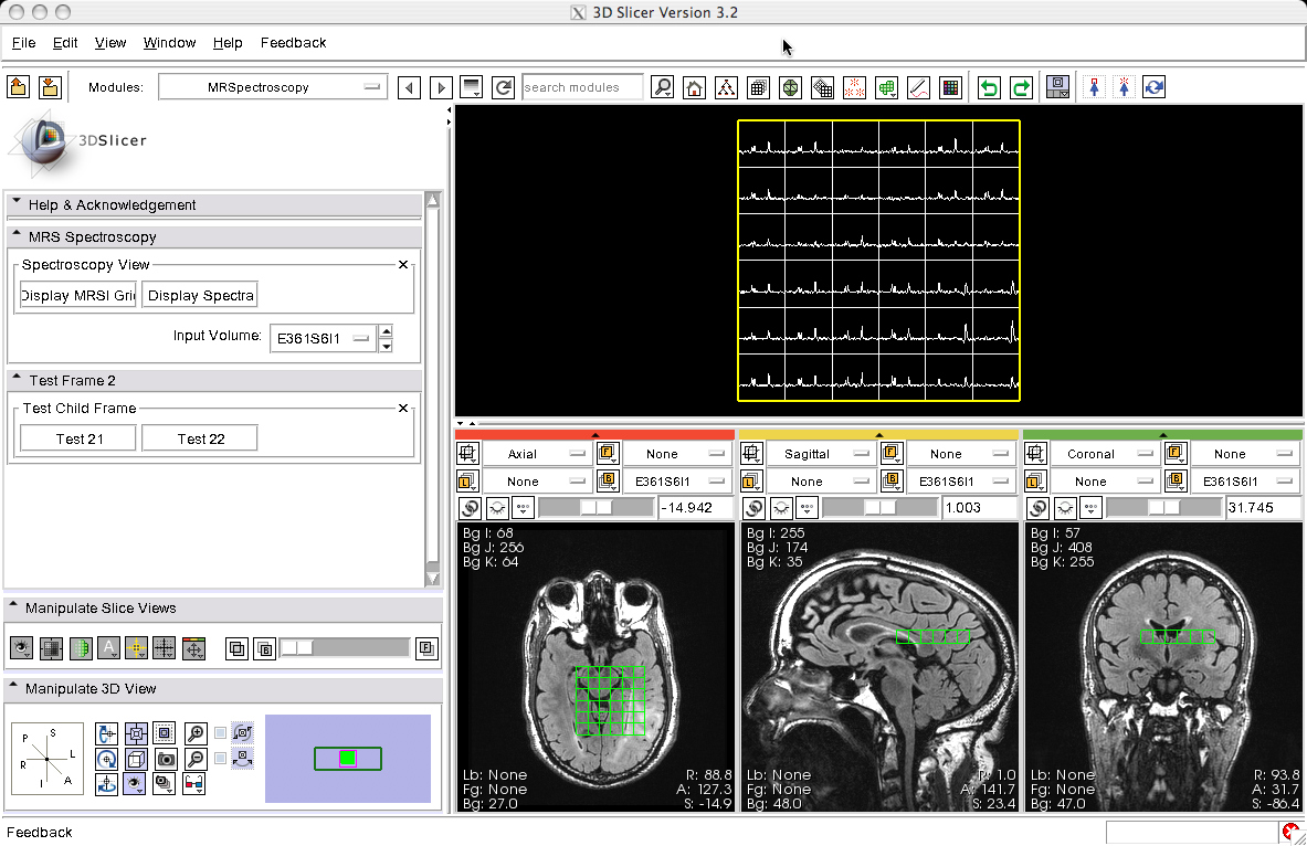
15 KB | A MRSI data processing module for Slicer | 1 |