File list
From NAMIC Wiki
This special page shows all uploaded files.
| Date | Name | Thumbnail | Size | Description | Versions |
|---|---|---|---|---|---|
| 22:09, 20 June 2010 | MBP-definition.jpg (file) | 16 KB | Definition of the median binary pattern feature. | 1 | |
| 21:56, 20 June 2010 | Masked-bkg mbp+2.png (file) |  |
79 KB | Distribution of MBPs shown with each pattern mapped to a unique color. | 1 |
| 21:55, 20 June 2010 | Masked-bkg mbp+2.tif (file) | 114 KB | Distribution of MBPs shown with each pattern mapped to a unique color. | 1 | |
| 20:42, 20 June 2010 | Mask 2TS0005 -1NU.png (file) | 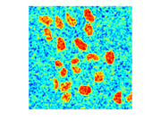 |
589 KB | MBP histogram of all non-uniform patterns. | 1 |
| 20:41, 20 June 2010 | MBP 2TS0005 17U.png (file) | 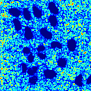 |
452 KB | MBP Uniform pattern 17 histogram | 1 |
| 20:41, 20 June 2010 | MBP 2TS0005 16U.png (file) |  |
436 KB | MBP Uniform pattern 16 histogram | 1 |
| 20:40, 20 June 2010 | MBP 2TS0005 15U.png (file) |  |
347 KB | MBP Uniform pattern 15 histogram | 1 |
| 20:40, 20 June 2010 | MBP 2TS0005 14U.png (file) |  |
324 KB | MBP Uniform pattern 14 histogram | 1 |
| 20:39, 20 June 2010 | MBP 2TS0005 13U.png (file) |  |
433 KB | MBP Uniform pattern 13 histogram | 1 |
| 20:38, 20 June 2010 | MBP 2TS0005 8U.png (file) | 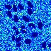 |
180 KB | MBP Uniform pattern 8 histogram | 1 |
| 20:36, 20 June 2010 | MBP 2TS0005 7U.png (file) | 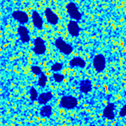 |
307 KB | MBP Uniform pattern 7 histogram. | 1 |
| 20:30, 20 June 2010 | MBP 2TS0005 6U.png (file) |  |
270 KB | MBP Uniform pattern 6 histogram. | 1 |
| 20:30, 20 June 2010 | MBP 2TS0005 5U.png (file) |  |
371 KB | MBP Uniform pattern 5 histogram. | 1 |
| 20:07, 20 June 2010 | FGPAC mask 101iter stack0001frame0005.png (file) | 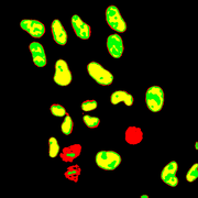 |
18 KB | Multiphase GPAC level set segmentation mask for HeLa cell cycle sequence 2TS frame 5. | 1 |
| 20:04, 20 June 2010 | 2TS0005 stack0001frame0005.png (file) | 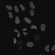 |
663 KB | HeLa cell cycle raw fluorescence image sample from sequence 2TS frame 5. | 1 |
| 19:33, 20 June 2010 | 2TS0005 stack0001frame0005.tif (file) | 1 MB | HeLa cell cycle raw fluorescence image sample from sequence 2TS frame 5. | 1 | |
| 16:16, 20 June 2010 | Zebrafish-nuclei-membrane-channel-multiphase.png (file) | 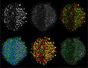 |
637 KB | Fused multiphase results for zebra fish dual channel fluorescence image slice. Images shown top to bottom include nuclei channel, membrane channel, RGB membrane-nuclear false color, RGB color of [nuclear, membrane enhanced saliency tensor, Mask-blobiness] | 1 |
| 15:15, 20 June 2010 | Histopathology GVD2 grade4 2 zoom.png (file) | 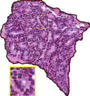 |
951 KB | Region segmentation and nuclei detection results for a sample Grade 4 image. Blue: Detected nuclei centers, Red: nuclei boundaries obtained using marker controlled watershed segmentation. Nuclei center recall:81%, precision:96%. | 1 |
| 15:14, 20 June 2010 | Histopathology mvls 4class.png (file) | 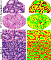 |
523 KB | Multiphase vector-based level sets (MVLS) segmentation of four tissue types into three categories: nuclei (red & black), lumen (green), epithelial cytoplasm (yellow). | 1 |
| 15:02, 20 June 2010 | MRI-multiphase-gpac.png (file) |  |
243 KB | MRI mulitphase GPAC segmentation using 4-phases. | 1 |
| 14:01, 20 June 2010 | Multiphase-GPAC-HeLa-segmentation.png (file) | 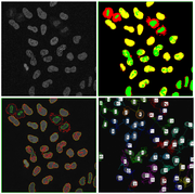 |
499 KB | Multiphase GPAC level set results for TS2 HeLa cell cycle sequence. | 1 |